Lateral Collateral Ligament Release in Fixation of Complex Distal Humerus Fractures
The authors describe a technique for extensile approach to the distal humerus via lateral collateral ligament release as an effective means of improving anterior articular visualization.
Case Overview: The patient is an elderly female with a distal humerus fracture after a fall. Imaging revealed a comminuted intraarticular distal humerus fracture involving the lateral column with coronal shear capitellar fragment. Open reduction and internal fixation was performed with olecranon osteotomy and lateral collateral ligament (LCL) complex release during exposure followed by dual plating.
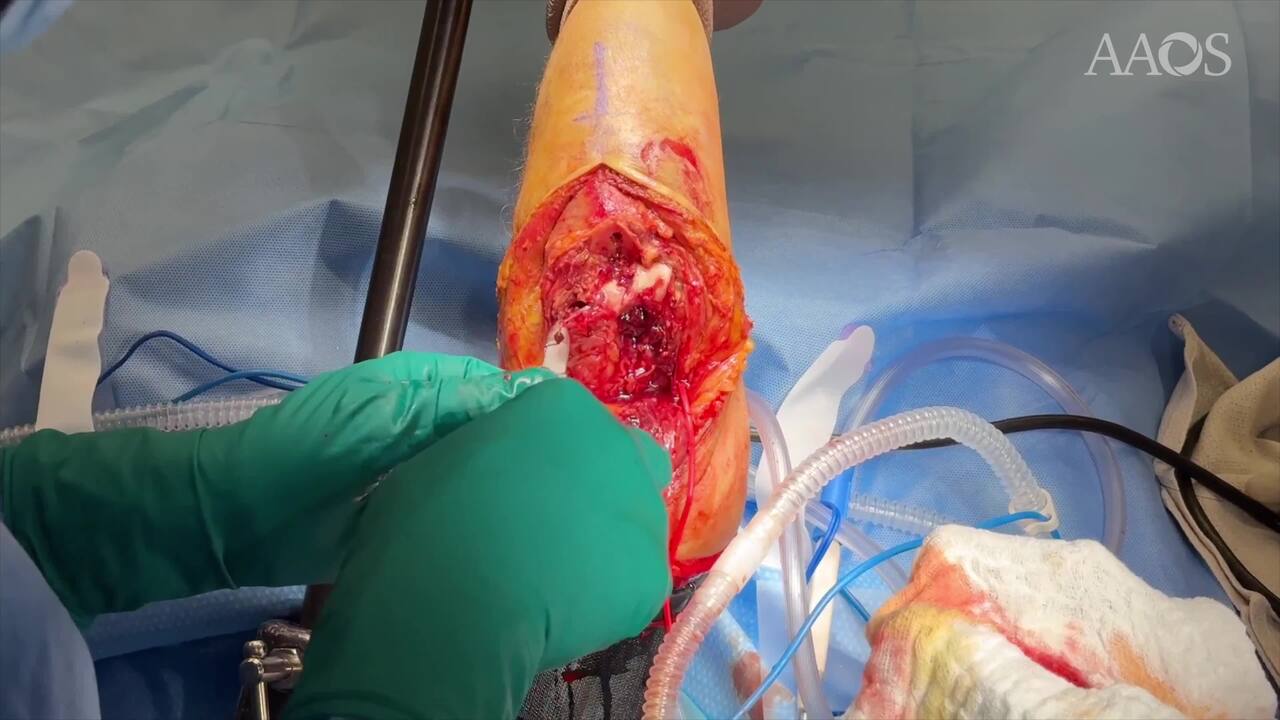
Method/Technique: The patient was placed supine on the operating table with the McConnell arm attachment. A posterior longitudinal incision was made and full thickness skin flaps were raised. Medially the ulnar nerve was identified and temporarily transposed. Olecranon osteotomy was performed and the triceps and olecranon were reflected posteriorly. Due to inadequate visualization of the anterior articular surface including the capitellar shear component, a lateral release was then performed. The LCL complex was tagged using two #2 Fiberwire sutures in locking Krackow fashion. It was then sharply released off the anterior surface of the lateral epicondyle. The joint was opened by hinging medially, exposing the entirety of the articular surface. Fracture reduction and fixation was then performed with perpendicular lateral plates and supplementary medial screw fixation. The construct was checked under fluoroscopy and direct visualization to confirm anatomic reduction and lack of intraarticular screw penetration. After fixation of the distal humerus, the olecranon osteotomy was reduced with intramedullary cannulated screw. The LCL complex was repaired to the lateral plate via the #2 Fiberwires. The ulnar nerve was returned to its native position. After closure the patient was placed into a long arm posterior splint to be worn for two weeks prior to beginning gentle range of motion guided by occupational therapy.
Results: In review of 12 patients treated with this technique, all were found to have satisfactory outcomes. No patients demonstrated fixation failure or physical exam consistent with elbow instability. Two patients required return to OR for post-traumatic heterotopic ossification excision and contracture release. During reoperation, the repaired LCL complex was intact and fully healed.
Summary: The authors describe a technique for LCL complex release following olecranon osteotomy to optimize anterior articular visualization of challenging distal humerus fractures. The LCL complex is sharply elevated during approach, then later repaired to its origin via two #2 Fiberwire Krackow sutures. This technique is safe and effective with good short term outcomes. Despite the concern for posterolateral instability, postoperative stiffness remains the most common sequelae during recovery.