Rotator Cuff Tear Patterns: Recognition and Treatment
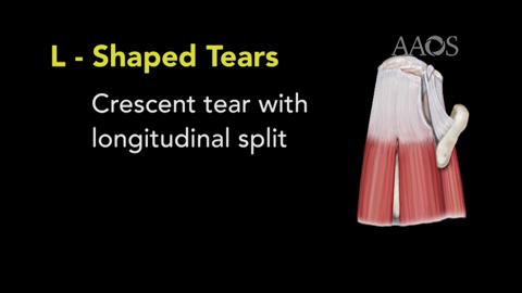
Over the past century, numerous classification systems have been developed to help describe tear morphologies and risk factors for tear progression or failure of arthroscopic repair using various two-dimensional image planes. Examples include the sagittal and coronal topographic characteristics described by Patte, the stages of muscle atrophy and fatty infiltration described by Thomazeau et al and Goutallier et al, respectively, and the classifications of tear severity described by both Ellman and Snyder et al. While each of these classification systems can still be useful for the description of specific features of rotator cuff tears, their collective ability to predict the appropriate repair technique and prognosis is very limited. With the advancement of arthroscopic surgery, investigators have organized rotator cuff tears according to their three-dimensional appearance and preferred technique for arthroscopic repair. The four most common tear patterns are crescent-shaped, U-shaped, L-shaped (or reverse L-shaped), and massive, contracted, immobile tears, each of which has a recommended technique for repair that is most likely to result in a successful outcome according to the best available clinical and biomechanical data. The purposes of this video are (1) to review the four most common tear patterns encountered in clinical practice, (2) to describe the recommended sequences of repair, and (3) to demonstrate the repair of each tear pattern using a case-based approach.