Posterior Hindfoot Needle Endoscopy in the Office Setting
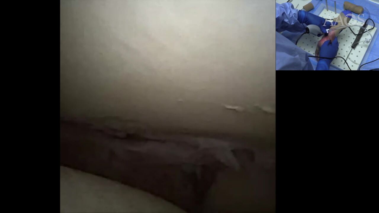
Background Posterior hindfoot disorders encompass a wide spectrum of pathologies, including posterior ankle impingement syndrome (PAIS), disorders of the flexor hallucis longus (FHL) tendon, osteochondral lesions (OCLs), and coalitions. Causes can be anatomic—such as a pathologic posterolateral talar process known as a Steida process or an os trigonum, related to overuse, such as seen in ballet dancers or soccer players—or post-traumatic in nature. Treatment of posterior hindfoot disorders was historically performed with open exploration; however, multiple complications were reported, including adhesions, symptomatic scar formation, wound breakdown, neurovascular injury, and stiffness. Studies involving the use of endoscopic techniques have shown lower complication rates, shorter recovery time, less blood loss, and less postoperative pain while maintaining comparable functional outcomes. Recent advancements in needle arthroscopy such as chip-on-tip image sensor technology have pushed the boundaries of minimally invasive surgery. This even allows procedures to be performed in the office. Purpose In this video, we describe the advantages and disadvantages of posterior hindfoot endoscopy and demonstrate the technique, including relevant anatomic landmarks, proper portal placement, and endoscopic resection. Methods This video presents first a cadaver demonstration, with relevant landmarks, including surface anatomy, as well as a endoscopic tour highlighting all relevant landmarks. After this, a case demonstration is presented for a patient with os trigonum. The presentation concludes with presentation of relevant outcome studies. Results The patient has significant improvement in range of motion and function after the procedure, without any complications. Conclusion Posterior hindfoot endoscopy is a safe, reproducible procedure that can treat a variety of posterior impingement disorders.