Instability and Osteoarthritis of the Sternoclavicular Joint: From Anatomy to Surgical Treatment
Introduction
The sternoclavicular joint is one of the most rarely injured joints in the body; however, it may become a source of symptoms secondary to traumatic dislocation, spontaneous instability in patients with generalized joint laxity, or osteoarthritis. Although uncommon, injuries to the sternoclavicular joint may be severe because of the proximity to posterior vascular structures; however, anterior dislocations are more common because of the greater strength of the posterior sternoclavicular ligament. Nonsurgical management commonly is considered a first-line treatment option for most patients, with closed reduction of the joint; however, continuous pain and recurrent instability after nonsurgical management are indications for surgical management. Surgical management of chronic instability and painful arthritis of the sternoclavicular joint includes resection of the medial end of the clavicle, achieving better results by performing tendon graft reconstruction of the sternoclavicular ligaments.
Purpose
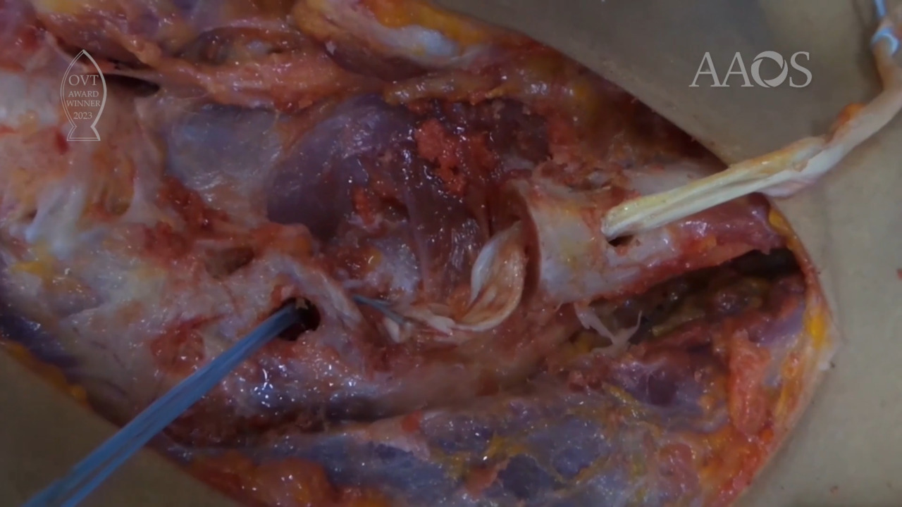
This video shows the normal anatomy of the sternoclavicular joint with its ligaments and demonstrates sternoclavicular joint reconstruction with the figure-of-eight with a semitendinosus tendon graft in a cadaver laboratory. In addition, the outcomes of surgical management via the described technique are reviewed.
Methods
The video shows the gross anatomy of the sternoclavicular joint with its ligaments and demonstrates sternoclavicular reconstruction with the figure-o-eight with tendon graft and sternoclavicular joint graft reconstruction via the sternal docking technique in a cadaver laboratory. Six patients with painful sternoclavicular joint instability and persistent pain in whom nonsurgical treatment failed are reviewed. All the patients underwent reconstruction of the sternoclavicular joint with the use of a semitendinosus tendon allograft via an anterior approach to expose the sternoclavicular joint along with the medial third of the clavicle. The sternoclavicular joint was disarticulated via subperiosteal dissection of the medial clavicle, and the intra-articular disk was removed. The retrosternal space was carefully dissected, and a curved periosteal retractor was placed retrosternally to protect the retrosternal structures before the bone tunnels were established. Figure-of-8 reconstruction was achieved by passing the allograft through two tunnels made in the sternum from anterior to posterior that are 4 mm in diameter and through two 4.0-mm tunnels drilled in the medial clavicle. Both ends of the graft were knotted together, and the construct was secured with the use of an additional nonabsorbable suture. The shoulder was immobilized in a sling for 6 weeks postoperatively. After 2 weeks, physical therapy was initiated, with pendulum and passive range of motion exercises performed until 6 weeks postoperatively (up to 90° of anterior elevation and abduction). Protraction and retraction of the scapula was avoided for 6 weeks postoperatively. Active-assisted range of motion was allowed at 6 weeks postoperatively, and strengthening exercises were initiated at 8 weeks postoperatively. Return to activities of daily living was possible 8 to 12 weeks postoperatively, and return to full contact sports activity was possible after 6 months postoperatively. The outcomes were assessed at a mean follow-up of 24 months (range, 12 to 48 months) via the Disabilities of the Arm, Shoulder and Hand score; the visual analog scale for pain; and patient satisfaction.
Results
Six patients (6 males) with a mean age of 25.3 years (range, 18 to 48 years; standard deviation [SD]± 11.32 years) were included in the study. All the patients had painful sternoclavicular joint instability with persistent pain in whom nonsurgical treatment failed. The mean Disabilities of the Arm, Shoulder and Hand score improved 15 points (range, 0 to 25; SD± 8.37); the visual analog scale for pain improved 1 point (range, 0 to 2; SD± 0.7); and patient satisfaction improved 9 points (range, 8 to 10; SD± 1) at final follow-up.
Conclusion
The painful sternoclavicular joint may be the result of traumatic dislocation and arthritic conditions. Reconstruction of the sternoclavicular joint with the use of tendon allograft results in good clinical outcomes.