Distal Biceps Tendon Repair Using a Single-Incision Dual-Anchor Technique
Background
Distal biceps tendon ruptures commonly present in middle-aged patients, resulting in deficits of strength and endurance. Surgical management results in improved functional outcomes; however, clear consensus on an ideal surgical treatment option does not exist. This video discusses the case presentation of a 45-year-old man who underwent distal biceps tendon repair via a single incision using a dual-anchor technique.
Indications
Distal biceps tendon repair is indicated in patients with clinical evidence and supporting MRI confirmation of a complete or partial rupture of the biceps tendon. Ideally, surgical treatment is performed within 1 to 2 weeks postinjury to minimize the amount of scar tissue and the severity of tendon retraction.
Technique Description
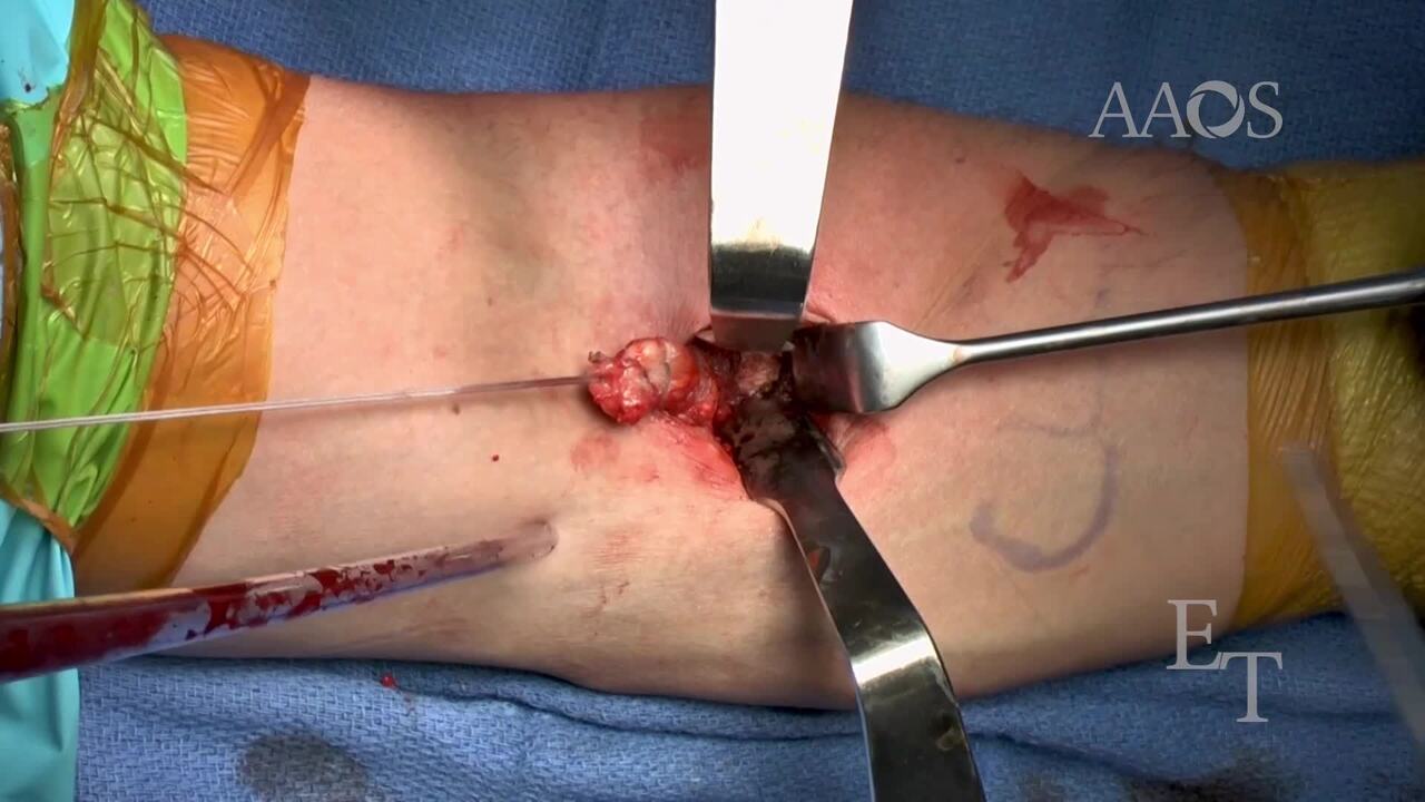
A single incision is created on the volar surface of the forearm approximately 15 mm distal to the main flexor crease. After dissection to and retrieval of the biceps tendon, a No. 2 FiberTag stitch (Arthrex) is placed distally and secured in a standard looped, locking fashion. An anchor is then placed in the most proximal aspect of the radial tuberosity and preliminarily placed around the tendon. An all-suture intramedullary cortical button is then placed in the distal aspect of the radial tuberosity. The FiberTag sutures are then shuttled through the button and tightened to anatomically reduce the distal biceps. The sutures on the proximal anchor are then used for supplemental fixation.
Results
An anterior, single-incision technique provides the exposure necessary for dual-anchor fixation and anatomic restoration of the distal biceps tendon. This approach results in improved flexion and pronation at 1 year postopertively, lower rates of heterotopic ossification, and lower rates of revision surgery.
Conclusion
Distal biceps tendon repair via an anterior, single incision affords excellent exposure for surgical repair. With a dual-anchor technique, the distal button allows for anatomic fixation while the proximal suture anchor provides secondary fixation and increases the bone-tendon interface.