Use of Patient-Specific Three-Dimensionally Printed Alignment Guides in Complex Revision Total Knee Arthroplasty
One of the most critical issues in knee revision surgery is the management of bone loss, especially in septic revision procedures. Lack of knowledge or mismanagement may compromise proper placement of new prosthetic components and affect the final clinical outcome. Therefore, accurate planning is essential.
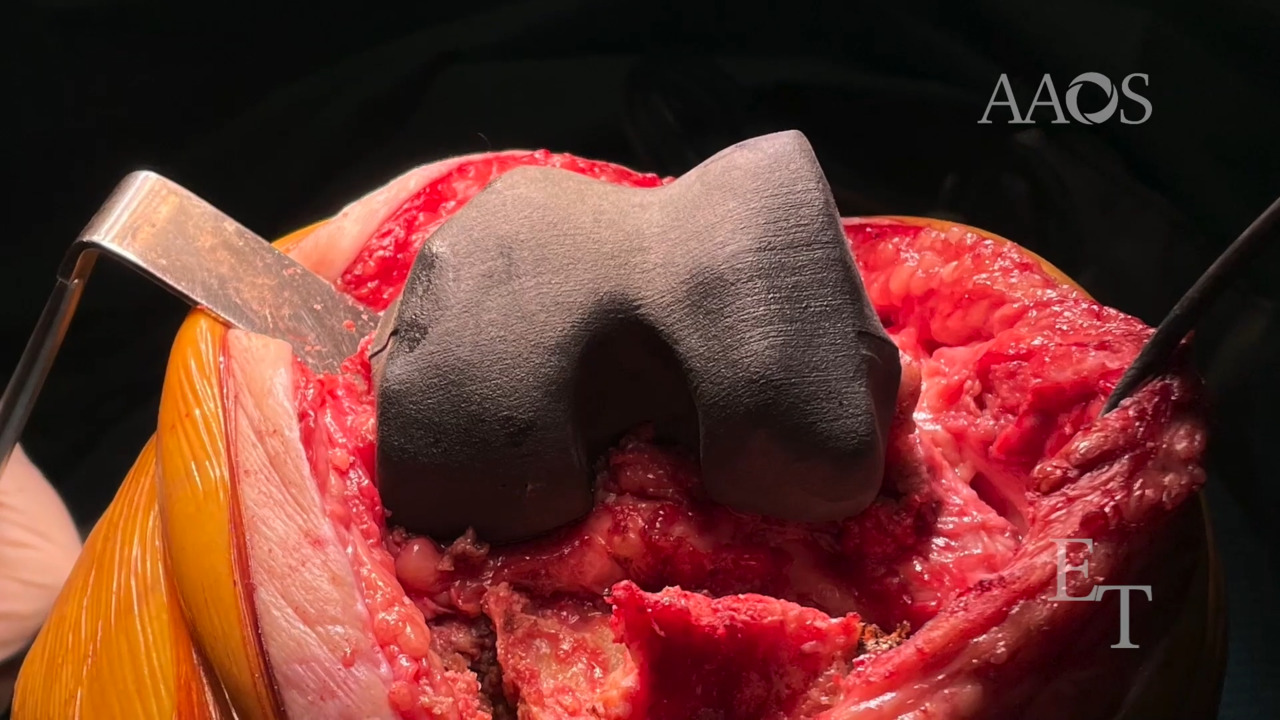
This video demonstrates an approach to complex two-stage knee revision that involves the use of patient-specific positioning guides, which can simulate bone loss and correct the positioning of revision prosthetic components. The first step involves obtaining a bilateral lower extremity CT scan after prosthetic explantation. The CT scans obtained are reprocessed using dedicated software. The three-dimensional reconstruction image of the two limbs is overlapped by mirroring with the contralateral limb to correctly estimate the size and shape of bone loss. On the reconstruction images of the femoral and tibial bone defects, markers are applied at the epicondyles and the trochlear groove for the femoral part and at the center of the tuberosity for the tibial part. The reconstruction images of the bone defects are then printed into three-dimensional models and used intraoperatively as trial components to fill bone loss and ensure restoration of gaps, correct sizing of the prosthetic components, and correct positioning of the prosthetic components in terms of alignment and rotation.