Endoscopic Shelf Acetabuloplasty Combined With Labral Repair and Cam Osteoplasty via Periportal Capsulotomy Access for Treating Patients With Acetabular Dysplasia
Introduction
Isolated hip arthroscopy generally is not accepted for the management of acetabular dysplasia. The authors of this video devised endoscopic shelf acetabuloplasty combined with labral repair and cam osteoplasty. A previous study reported on a series of 36 hips in which this procedure was performed. Patient-reported outcome measures substantially improved from preoperatively to postoperatively, and 90% of active patients returned to their preinjury activity level. Another study on a series of 23 artistic dancers reported that patient-reported outcome measures substantially improved, and 87% of the patients returned to dance at a mean of 9 months postoperatively.
Indication and Algorithm
Patients with borderline developmental dysplasia of the hip (lateral center edge angle of 20° to 25°) who have an anterior center edge angle less than 20°, a Tönnis angle greater than 15°, and a normal Shenton line are indicated for endoscopic shelf acetabuloplasty combined with labral repair, cam osteoplasty, and capsular repair. Patients with flank hip dysplasia (lateral center edge angle <20°) who have have no Shenton line breakage, a lateral center edge angle greater than 5°, a femoral neck-shaft angle less than 139°, and normal femoral anteversion are indicated for this procedure.
Contraindication
Contraindications include the presence of osteoarthritis (including severe cartilage damage) at the time of surgery, severe hip dysplasia (lateral center edge angle <5°), a broken Shenton line, a femoral neck-shaft angle greater than 140°, and femoral anteversion greater than 40°.
Surgical Technique
Portals: The anterolateral portal, mid-anterior portal, and proximal mid-anterior portal are made.
Shelf graft preparation: An autologous bone graft is harvested from the ipsilateral iliac crest. The graft donor site should be 2 cm posterior to the anterior superior iliac spine to avoid lateral femoral cutaneous injury. A 1.8-mm Kirshner wire is used to make 1.8-mm diameter drill holes. Two 1.5-mm Kirschner wires are introduced in these drill holes.
Intra-articular procedure: A 70° arthroscope is introduced through the anterolateral portal. A radiofrequency ablation device is then used in the anterolateral portal to further dilate the capsular opening. Dilation is then performed with the mid-anterior portal. The dilation width is 8 to 10 mm. The arthroscope is then reintroduced into the anterolateral portal. An 8.0-mm disposable cannula is placed through the mid-anterior portal. An anterosuperior tear is observed. Labral repair with suture anchors is then performed. After the central compartment procedure is completed, traction is released, and the cam lesion is exposed. After traction is released, Minitape is used to hang up the capsule, widening the space between the cam lesion and capsule. Viewing the mid-anterior portal, a burr is introduced in the mid-anterior portal without a cannula. Cam osteoplasty is then performed.
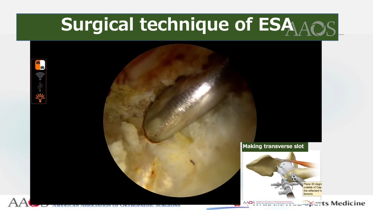
Shelf procedure: A 30° arthroscope is positioned in the extracapsular space. The soft tissue surrounding the reflected head is débrided with the use of a shaver and a radiofrequency ablator until the reflected head is visible. The four 2.3-mm guidewires are introduced using the parallel drill guide. Over-drilling is performed with the use of a 5.0-mm drill for each pin to create a slot, which is enlarged with the use of a shaver and osteotome. The optimal width and depth are then confirmed with the use of a dilator. Finally, shelf autograft is inserted into the slot with the use of skewer wires and press-fit into position with the use of cannulated bone tamps. Corticocancellous bone chips are packed above the new shelf.
Conclusion
Endoscopic shelf acetabuloplasty combined with labral repair and cam osteoplasty via periportal limited capsulotomy with repair is a less invasive and promising procedure for the treatment of patients with hip dysplasia.