High Tibial Osteotomy and ACL Reconstruction with Patient Specific Instrumentation
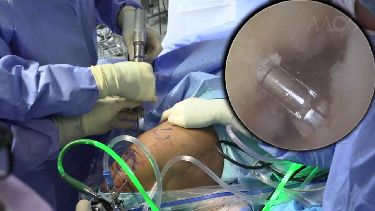
Background: High tibial osteotomy (HTO) has become a powerful tool to improve knee stability and alignment by correcting both coronal and sagittal plane deformities about the knee. When planning for an ACL reconstruction, deformity correction must be considered in addition to ACL graft choice, as previous studies have shown that varus alignment may negatively impact outcomes after ACL reconstruction due to increased ligament strain. In the setting of varus malalignment and ACL deficiency, the use of patient-specific guides can help overcome the technical challenges of surgically addressing multiplanar corrections, while simultaneously offering the benefits of less radiation, less operative time, and elimination of screw or tunnel convergence.Purpose: Through a case presentation, this video demonstrates the surgical technique for medial wedge opening osteotomy and bone-tendon-bone (BTB) autograft ACL reconstruction using patient-specific instrumentation (PSI) for patients with both knee instability and varus malalignment.Methods: Evaluation, diagnosis, and treatment of knee instability and varus deformity with medial opening wedge osteotomy and ACL reconstruction are illustrated through a case presentation of an active, young 27-year-old male patient with a chronic ACL tear associated with medial compartment arthritis.Results: Using PSI, intraoperative fluoroscopy demonstrated correction as expected from the preoperative plan. At the 6 weeks postoperative visit, our patient’s x-rays demonstrate intact hardware and interval healing at the osteotomy site. He is doing well, with minimal pain and improving knee ROM 0-110. While follow-up is needed on our patient, he is progressing through a standardized rehabilitation protocol.Conclusion: Digitally planned and executed HTO with ACL reconstruction using PSI is a promising tool to simplify and more accurately correct knee instability and malalignment, with additional benefits of shorter operative times and decreased radiation compared to traditional techniques. Early results show good radiographic and clinical outcomes.