The Extensile Posterior Approach for Complex Tibial Plateau Fractures
Introduction: Operative treatment of complex tibial plateau fractures requires an appropriate approach to not only visualize but also stabilize each fracture component at its apex. For posterior, posterolateral, and posteromedial-based fractures, the ideal implant would capture/buttress multiple small fracture fragments and extend close enough to the articular surface when needed. In this video, we present the extensile posterior approach for operative reduction and internal fixation of posteromedial and posterolateral plateau fractures in a 46-year-old male who presented with a multi-fragmentary tibial plateau fracture (AO/OTA 41C3.3) after a motor vehicle collision. This fracture seen here has a significant component that is posteriorly based, necessitating a posterior approach in order to control, reduce, and stabilize the posteromedial and posterolateral plateau fragments. It is anticipated that additional anteromedial and anterolateral approaches will be necessary to manage the anterior medial and lateral fracture components.
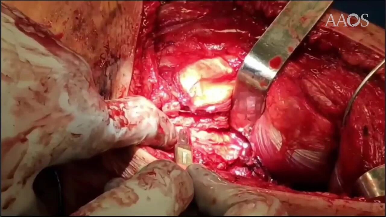
Surgical Technique: The patient is placed prone on a radiolucent table. After prepping and draping, a curvilinear, S-shaped posterior knee incision is made. The incision is carried down through the skin, extending proximally and laterally if an extensile dissection is needed. Dissection is carried down through the deep subcutaneous tissues down to the fascia overlying the posterior compartment. The tourniquet is placed but is not inflated. This is our preference so that if a vascular injury occurs, it can be immediately recognized. Once the deep fascia is divided, the medial head of the gastrocnemius is identified beneath the deep fascia, and dissection is performed to visualize the origin of the medial gastrocnemius. This can generally be done bluntly. Once the muscle belly is elevated, the tendon at the origin of the medial gastrocnemius is generally easy to identify and can be divided under direct visualization. Great care is taken not to extend the division of the tendon too laterally, to avoid endangering any deep neurovascular structures. Once the origin is divided, the muscle belly can be retracted laterally, giving an extensile exposure to the posterior tibia. The origin of the soleus is then identified and divided along the medial border of the tibia. Dissection is then carried laterally to get to the deep bony structures. At this point, the posterior medial fragment and its apex are well visualized. Pins can be placed distally, and retractors can be placed beneath the muscle mass proximally to allow visualization all the way to the posterior lateral plateau.
Deep dissection can be performed directly on the bony fragments, but great care must be taken to avoid dividing the insertion of the posterior cruciate ligament. The posterior medial fragment component is freed of soft tissues and the tibial shaft is manipulated with longitudinal traction with the knee in full extension to allow for reduction of the posterior medial fragment. This is done after interposed hematoma and soft tissues have been removed.
Once the medial side is reduced, it is held provisionally with K-wires, and fluoroscopy is used to confirm the reduction of the posterior medial fracture component. An anti-glide plate is contoured and placed at the apex posteromedially. In general, a mini-fragment plate provides adequate stability for these relatively small fragments. Non-locked screws are generally used in the shaft, and proximally either locked or non-locked screws can be used. A second plate can be placed more medially to provide additional stabilization on the medial aspect of the posterior medial fracture component. Fluoroscopy confirms reduction posteromedially, and attention is turned to the posterior lateral joint. With the gastrocnemius muscle retracted, the depressed segment of the posterior lateral joint can be directly visualized. In the presented case, the posterolateral fragment is manually reduced and supported with the use of biological cement. Note also that in this case, no direct fixation is placed into the posterior lateral component. Repair of the medial head of the gastrocnemius is performed with a simple figure of eight sutures, and the skin and deep soft tissues are closed in standard fashion.
Further Management in the presented case, a second anterior approach is necessary to complete the fixation of this very committed fracture with both anterolateral as well as anteromedial fixation.
Postoperative care: In general, the patient is immobilized in a hinged knee brace for 6 weeks. Active range of motion begins at two weeks postoperative. Given the fracture complexity, partial weight-bearing does not start till 8-10 weeks and then progresses to full weight-bearing over the following 6-8 weeks.