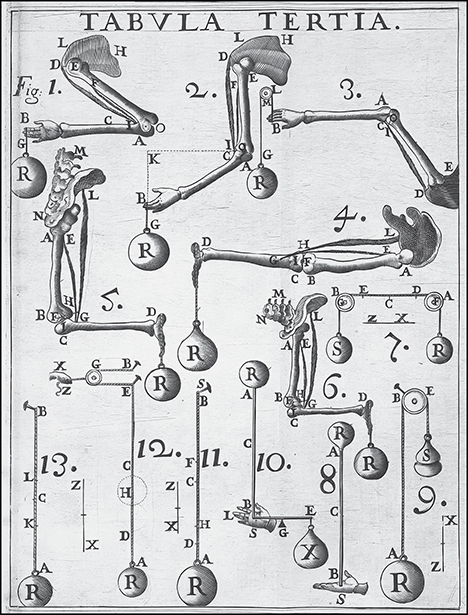
Fig. 1 Movement of human appendages and pulley systems, compared with principles of mechanics and statics by Giovanni Borelli
Source: Library of Congress, LC-USZ62-95253
Published 4/26/2024
|
Stuart A. Green, MD, FAAOS
Leonardo da Vinci (1452–1519), one of the first artists to study human cadavers, recognized that mechanical principles provide the basis for human movements. In his book A Treatise on Painting, Da Vinci wrote, “The science of mechanics is so noble and useful in comparison with all other sciences, that it is possible that all living organisms having the possibility of moving are governed by its laws.”
Since the beginning of recorded history, great minds have puzzled over limb movement and locomotion. Questions about these subjects appear in Egyptian hieroglyphic papyri. Socrates and Plato established a method of inquiry that encouraged others to seek explanations for processes observed in everyday life.
Aristotle (384–322 BC) wrote De Motu Animalium, a treatise explaining how humans and animals move. He concluded that an inborn spirit, the pneuma, caused muscles to expand and contract, resulting in bodily movement.
Galen of Pergamon (129–216 AD) was a prolific writer whose hundreds of treatises influenced medical thinking for the next 1,400 years. He subscribed to and amplified Aristotle’s four-humor concept of health and illness, along with a version of the pneuma concept. In De Usu Partium (On the Usefulness of Parts of the Body), Galen explained how the in-and-out movement of a vital fluid was responsible for muscle action. After all, one could see that a leather sac would widen and shorten when filled with fluid and would move in the opposite direction when that fluid was withdrawn. He concluded that nerves conducted the fluid to and from muscles, having observed that cutting an innervating nerve paralyzed that animal’s muscle. His perspective influenced European and Arabian thinkers until his ideas were overthrown by Renaissance investigators, including William Harvey (1578–1657), author of De Motu Cordis.
Galileo Galilei (1564–1642) also contributed to understanding the mechanics of living systems, as did anatomist Andreas Vesalius (1514–1564). However, Giovanni Alfonso Borelli (1608–1679) can rightfully be considered the father of biomechanics. He realized that muscles can only contract; thus, they shorten actively. He concluded that muscle elongation is a passive process, recognizing, for instance, that expiration occurs through elastic rebound. He gave his masterwork, De Motu Animalium, the same title as Aristotle’s earlier effort.
Borelli documented that bones are levers that increase the range of motion of terminal body parts, compared to the excursion of the muscle generating the motion (Fig. 1). For this reason, Borelli determined that muscles exert great force during contraction over a short range, which the skeletal arrangement converts to a lesser force of greater range. For instance, the end of the biceps tendon inserting on the radial tuberosity moves about 3 inches to cause the palm to traverse an arc 30 inches long. This 10-to-1 ratio implies that, to lift a 10-pound weight, the biceps must exert 100 pounds of force on the tuberosity.
The fluid theory of muscle movement was finally zapped by Luigi Galvani (1737–1798), who demonstrated that electrical current, rather than fluid flow, makes muscles move. Popular demonstrations of this phenomenon followed, including the electrification of recently deceased cadavers. Presumably, such displays inspired Mary Shelley to write Frankenstein.
Modern advancements
Steady progress in biomechanics followed thereafter. For instance, Ernst, Wilhelm, and Eduard Weber located the center of mass of the body in 1836 and recognized that this point oscillates through space and time with forward locomotion.
With the advent of photography, Eadweard Muybridge and Étienne-Jules Marey developed techniques to document the motion of humans and animals in ways never before thought possible. This led to further advances in biomechanics by multiple researchers, including the evolution of force plates in 1938 and dynamic electromyography.
With the availability of these recording techniques, Verne T. Inman, MD, PhD, at the University of California, San Francisco, along with colleague J.B. Saunders and University of California, Berkeley, engineer Howard Eberhart, created a gait laboratory to study how amputees walk while wearing prostheses of different designs. They employed cine-photography, implanted electromyography electrodes, a floor-mounted force plate, and other features familiar to anyone studying human locomotion today. They even screwed protruding orthopaedic pins into their subjects’ bones to improve analysis.
The first textbook about biomechanics written by an orthopaedic surgeon was Mechanics of Normal and Pathological Locomotion in Man, published in 1935 by the University of Iowa’s Arthur Steindler, MD. Dr. Steindler’s book is filled with diagrams demonstrating the lever action of muscles, electromyography tracings in normal and pathologic conditions, and photographs of various individuals with deformities that affected their frontal and sagittal balance. Dr. Steindler founded the university’s Department of Orthopaedic Surgery and was responsible for training faculty colleague Ignacio Ponseti, MD, and hundreds of other prominent orthopaedic surgeons, including Ruth Jackson, MD, the first female board-certified orthopaedic surgeon.
Viennese by birth and educated in Europe, Dr. Steindler’s initial job after emigrating to the United States was as the assistant to John Ridlon, MD, at Chicago’s Home for Crippled Children. (Dr. Ridlon had spent time in Liverpool, studying with Hugh Owen Thomas and his nephew, Robert Jones, the founders of orthopaedic surgery as a distinct medical specialty.) Dr. Steindler next moved to Des Moines, Iowa, and from there to Iowa City to become the first orthopaedic surgeon at the University of Iowa.
Another influential textbook, Orthopaedic Biomechanics, was authored by Victor Frankel, MD, PhD, an orthopaedic surgeon trained at New York’s Hospital for Joint Diseases, with additional education at the University of Uppsala in Sweden, where he earned a PhD in biomechanics. In writing the 1970 monograph, Dr. Frankel teamed up with engineer Albert Burstein, PhD, when they worked together at Case Western Reserve University in Cleveland.
Their book goes beyond Dr. Steindler’s work by including mechanical engineering principles (and mathematical formulae) applied to bones and joints, along with information about how viscoelastic tissues (i.e., ligament, tendon, cartilage) behave under load. Other topics discussed in the book include joint lubrication and kinematic principles. The book ends with a discourse on implant design and failure.
During the past half-century, many departments of orthopaedic surgery have added biomechanics laboratories and engineers to their endeavors. The teaming of clinical scientists with engineers has resulted in remarkable advances in the care of patients with musculoskeletal disorders and diseases.
This issue of AAOS Now contains articles about many aspects of applied biomechanics, including the kinesiology of normal and abnormal human gait, techniques and devices to increase athletic performance, the development of advanced prosthetic designs, the testing of orthopaedic devices before marketing to the public, and advances in biomaterials research.
Stuart A. Green, MD, FAAOS, is cofounder and past president of the Limb Lengthening and Reconstruction Society; past president of the Association of Bone and Joint Surgeons; and a clinical professor of orthopaedic surgery at the University of California, Irvine. He is the son, first cousin, and father of AAOS Fellows. Dr. Green is a member of the AAOS Now Editorial Board.