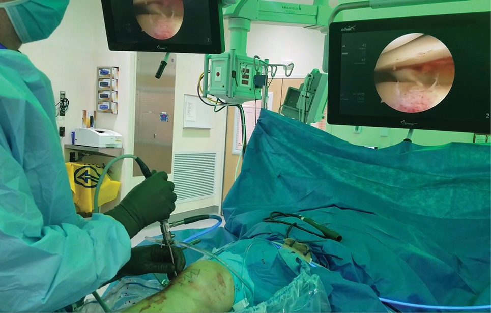
Figure 1: Arthroscopic visualization of the tibial plateau fracture
Published 12/17/2025
|
Stuart J. Fischer, MD, FAAOS
Editor’s note: The following article is a review of a video available via the AAOS Orthopaedic Video Theater (OVT). AAOS Now routinely reviews OVT Plus videos, which are vetted by topic experts and offer CME. For more information, visit aaos.org/OVT.
Tibial plateau fractures are a common articular injury accounting for 1% to 2% of all fractures. As with any articular injury, careful reconstruction of the joint surface will lead to a better result. Many articular fractures, such as those in the wrist, elbow, ankle, and knee, are being treated with a less invasive, arthroscopically assisted technique. This allows for better visualization of the joint surface as well as smaller incisions with less dissection.
A recent AAOS OrthoDome® video features a group of orthopaedic surgeons at the University of Minnesota demonstrating their technique for arthroscopically assisted lateral tibial plateau fixation. They emphasize that this technique is suitable for acute isolated lateral tibial plateau fractures with a restorable cortical envelope. This technique may not be appropriate for fractures with lateral cortical comminution, medial extension, or major soft-tissue injuries.
The video demonstrates the case of a female patient, aged 40 years, who sustained injury to her right leg as the result of a motor vehicle accident. The procedure was performed in an ambulatory surgical center. Preoperative CT templating was done to determine the position of a medial cortical window and direction of a bone tamp used to elevate the depressed articular surface.
The patient is positioned supine and draped for an arthroscopic knee procedure. The authors use a femoral distractor but note that this is optional.
The arthroscope is inserted through an anterolateral portal that is used for viewing the articular surface (Figure 1). A medial cortical window is then created with a tamp and a mallet to reduce the fracture. The fracture is elevated and reduced under fluoroscopy, and bone substitute is injected through the medial cortical window. Fracture alignment is confirmed by arthroscopy.
A small incision is made laterally. Guidewires are then inserted under fluoroscopic control. First, a partially threaded cancellous screw is inserted to achieve compression. Two additional fully threaded cancellous screws are used to complete the fixation. The reduction is viewed arthroscopically once more, and the wounds are closed.
During the procedure, pressure measurements are taken in the leg to make sure there are no signs of impending compartment syndrome. For most of the procedure, the soft-tissue pressures are less than 30 mm of mercury. The authors recommend that, if possible, the patient’s diastolic blood pressure be kept in the range of 90 mm of mercury.
After surgery, the knee is placed in a hinged brace and range of motion exercises are started. The patient is kept non-weight-bearing for six weeks. Aspirin is given for deep vein thrombosis prophylaxis.
The video also includes a review of other literature about arthroscopic tibial plateau fixation. One study indicated shorter tourniquet time as compared to a standard procedure and less time for the patient to advance to full weight-bearing. Most importantly, this study showed reduced joint space narrowing. Another study observed less blood loss, shorter surgical time, and better postoperative range of motion with an arthroscopic procedure.
The literature also indicated higher rates of surgical complications in traditional surgery, along with a greater incidence of medical complications and postoperative ED visits.
The authors stated that their technique provides the benefit of “direct visualization of the joint space with minimal dissection.”
Overall, this video provides a useful, easy-to-follow demonstration of the procedure for trauma surgeons and general orthopaedic surgeons who treat lower-extremity fractures.
Stuart J. Fischer, MD, FAAOS, is an orthopaedic surgeon in private practice in Watchung, New Jersey. He also serves on the AAOS Committee on Ethics and Outside Interests, AAOS Adult Reconstruction — Hip Program Committee, and the AAOS Digital Health Task Force. Dr. Fischer is also a member of the AAOS Now Editorial Board.
Video details
Title: Arthroscopic-Assisted Lateral Tibial Plateau Fixation with Percutaneous Screws
Authors: Mai P. Nguyen, MD, FAAOS; Erik A. Lund, MD, FAAOS; Emily H. Naclerio, MD; Marc F. Swiontkowski, MD; David Weatherby, MD, FAAOS, FRCSC; Jessica Lu Xu, MD, MS
Published: March 01, 2025
Time: 8:59
Tags: Trauma, Tibial Plateau Fractures, Fixation
Visit aaos.org/OVT to view this award-winning title and more than 1,600 other videos from across orthopaedic topics, institutions, practice management, and industry.