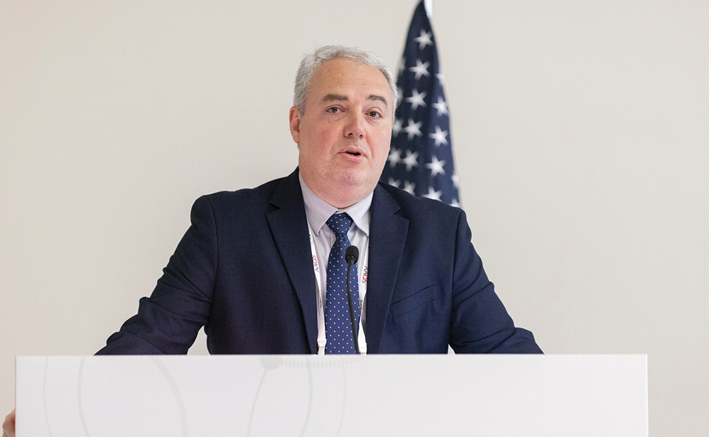
Shafic A. Sraj, MD, MBA, FAAOS, moderated a panel discussing various tools surgeons can utilize to support the evaluation and management of musculoskeletal conditions in the office setting.
Published 12/17/2025
|
Leah Lawrence
High-frequency, nonaudible sound — ultrasound — has only been harnessed by medicine in recent history. In modern orthopaedic practice, ultrasound has great appeal, according to Shafic A. Sraj, MD, MBA, FAAOS.
“Ultrasound is a powerful tool. You can use it in the office; it is on-demand, portable, and very quick,” said Dr. Sraj, associate professor in the Department of Orthopaedics at West Virginia University. “It doesn’t require prior authorization and is really cheap.” Ultrasound is empowering for orthopaedic surgeons because it combines the skills of three people into one technology: a technician, a radiologist, and a surgeon.
At the AAOS 2025 Annual Meeting, Dr. Sraj moderated Symposium J, “Office orthopaedics: Injectables, ultrasonography — new frontiers.” The session explored various tools surgeons can utilize to support the evaluation and management of musculoskeletal conditions in the office setting, including several presentations on the benefits and logistics of integrating ultrasound technology into clinical care.
Harnessing ultrasound
Dean W. Ziegler, MD, FAAOS, associate clinical professor at the Medical College of Wisconsin Department of Orthopaedic Surgery, encouraged session attendees to embrace ultrasound. “Look at ultrasound as increasing the value of your practice,” he said. In the right hands, this technology can allow orthopaedic surgeons to increase quality without markedly increasing cost, and to decrease cost without compromising quality.
As an example, Dr. Ziegler discussed a comparative study he performed 20 years ago looking at in-office ultrasound to evaluate integrity of the rotator cuff and comparing the imaging results with findings at surgery. Ultrasound was effective for imaging the rotator cuff with sensitivity, specificity, positive predictive value, and negative predictive values of 94.1%, 96.1%, 96.6%, and 93.2%, respectively, for partial-thickness tears.
“The key is technique,” Dr. Ziegler said. “The surgeon is the method.”
When using ultrasound, Dr. Ziegler still takes a thorough history, performs a physical exam of the affected shoulder first, and obtains plain radiographs. His indications for ultrasound are objective findings of cuff pathology on the physical exam and a patient who has failed appropriate nonoperative management. “I have found that ultrasound is a very effective and efficient approach to management of musculoskeletal disorders,” Dr. Ziegler said.
He added that to get the best ultrasound images, surgeons must have a great 3D understanding of the anatomy being evaluated. He noted that ultrasound requires some of the same skills as arthroplasty: Move the probe, rotate the probe, move the structure. Be comprehensive and consistent.
Dr. Ziegler closed by saying that it is important to remember that although ultrasound can make a structure appear worse than it is, it can never make it appear better than it is.
“Keep looking at the structure,” he said. “You are going to see it as good as it’s going to get.”
Interventional applications
Next, Christopher M. Jobe, MD, FAAOS, professor of orthopaedic surgery at Loma Linda University School of Medicine, walked attendees through some interesting interventional cases where he was able to successfully use ultrasound, including the removal of a foreign body — the back end of a shotgun shell — from a patient’s elbow.
Using case images and videos, Dr. Jobe demonstrated how ultrasound allowed him to assess the position of the foreign object, the damage it had caused, and nearby vessels. He used biplanar localization to retrieve the foreign body along the path of destruction it caused. Dr. Jobe also demonstrated the use of ultrasound in a frozen shoulder case, for the diagnosis of infection in a swollen shoulder, and to guide the injection of a biologic.
Why not ultrasound?
Finally, Robert Campbell Vercio, MD, of Puget Sound Orthopaedics, closed out the ultrasound presentations with some details about how to incorporate the technology into practice. When Dr. Vercio joined a private practice after completing his training, there was no ultrasound. He knows he is not alone in this situation.
“A lot of groups say they don’t want to spend the money [on ultrasound],” Dr. Vercio said, acknowledging that it can be challenging to get started. Typically, he has found that advocates for the use of ultrasound will run into three arguments: Surgeons do not have ultrasound, they do not know how to use it, or they are worried it will slow them down.
Available ultrasound technology can be grouped into three categories, with differing price tags and capabilities. The first type is a point-of-care ultrasound device, which typically costs around $4,000. This type of device is inexpensive, Dr. Vercio explained, pulling his own point-of-care ultrasound device from his pocket. It can have a small screen when connected to a phone but can also connect with an iPad or tablet.
“These are not going to give the highest quality, but it gets you started,” he said. “It opens the door.”
The next category is a portable ultrasound machine, or a “briefcase” model, which has a price tag between $10,000 and $20,000. With that price comes a significant step up in quality.
“You get better probes, and that opens the door to more techniques,” Dr. Vercio said.
The last category is a large ultrasound machine typically seen in hospitals, which costs upward of $80,000 and is not typically seen in private practice.
Dr. Vercio said that ultrasound procedures are typically reimbursed via three main codes: 76881 for complete diagnostic ultrasound, 76882 for limited diagnostic ultrasound, and 76942 for ultrasound guidance for needle placement.
“You do need to document your use. If you do one code five days a week, you are going to generate enough to pay for an ultrasound,” he said.
Obtaining a machine is the first step to getting better at using ultrasound. Another benefit of a small and portable ultrasound machine is that surgeons can practice on friends, family, or themselves, Dr. Vercio noted. Surgeons can also take a class or buy a book on ultrasound.
Finally, he emphasized that the use of ultrasound should not slow down workflow. In fact, it can serve as a tool to help distract patients during injections.
“You can do this,” Dr. Vercio said. “There is no reason not to!”
Leah Lawrence is a freelance writer for AAOS Now.
Reference
- Ziegler DW. The use of in-office, orthopaedist-performed ultrasound of the shoulder to evaluate and manage rotator cuff disorders. J Shoulder Elbow Surg. 2004;13(3):291-7. doi: 10.1016/j.jse.2004.01.017.