

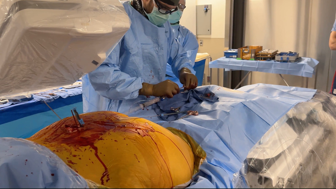
The surgical team fills the photodynamic nail under fluoroscopy.
Published 7/28/2026
|
Odion Binitie, MD, FAAOS
At a Glance
- Photodynamic nails offer a minimally invasive option for stabilizing impending pelvic fractures caused by metastatic disease.
- Combining photodynamic nails with cannulated screws may provide durable fixation while facilitating a rapid return to systemic cancer treatment.
- Successful implementation requires careful patient selection, advanced imaging, and close collaboration with interventional radiology.
Estimated read time: 3 minutes
Editor’s note: The following article is a review of a video available via the AAOS Orthopaedic Video Theater (OVT). AAOS Now routinely reviews OVT Plus videos, which are vetted by topic experts and offer CME. For more information, visit aaos.org/OVT .
Photodynamic nails (PDNs) increasingly are being used in oncology and trauma. These systems are indicated for the treatment of traumatic, pathologic, or fragility fractures in long bones. The technique involves the percutaneous insertion of a small flexible tubular balloon, which is then filled with a liquid monomer that expands to conform to the bone. The polymer is then hardened by visible LED blue light, curing it in minutes to a rigid, load-bearing implant.
Alan Woolridge, MD, MPH, FAAOS, and colleagues present an OVT on PDN use in metastatic pelvic and sacral lesions. The authors begin with background information on conventional interventions for impending pathologic fracture and the advantages of minimally invasive procedures that allow patients to return to systemic therapies quickly. Indications in the pelvis are minimally displaced or impending pathologic lesions, and contraindications are maximally displaced fractures or protrusio.
The case shown was a 48-year-old female patient with metastatic breast cancer with expansile lytic lesions in the right ilium and sacrum with nondisplaced fractures. She continued to have pain after chemotherapy and radiation. Preoperative planning was performed based on a CT for the planned trajectory of screws and the PDN. The procedure was performed in the interventional radiology suite with the use of augmented fluoroscopy. The patient was placed prone and a trocar was introduced into the posterior ilium, into which a guide wire was passed.
---
Related Content
- How safe are minimally invasive photodynamic intramedullary implants? (AAOS Now)
- Demystifying tumor classifications: key changes in bone and soft-tissue tumors (AAOS Now)
- Musculoskeletal Tumor Registry (MsTR) Annual Report (MsTR)
---
A second trocar was placed into the S1 corridor. Both screw paths were reamed under fluoroscopy, and a 3D spin was performed to confirm placement. A cannulated S1 screw was then placed. The corridor for the PDN was reamed over a guidewire, and it was inserted. The S1 screw was backed up to be later passed into the PDN. The PDN balloon was inserted through a sheath. The PDN was then filled with the monomer, filling portions of the lesion (Figure 1). The LED light was applied to harden the monomer, which can take 10 to 15 minutes depending on the size of the implant. Once hardened, the sheath is removed. The two screws were then advanced to interdigitate with the PDN. A final 3D spin confirmed appropriate placement.
Pearls of wisdom from the video include a multidisciplinary team approach with interventional radiology and the use of advanced imaging through a minimally invasive approach. Various interventions can be performed in addition to a PDN, including embolization, ablation, and cement. Pitfalls include it being a new procedure without significant data and it being most successful when performed in conjunction with interventional radiology.
Odion Binitie, MD, FAAOS, is a professor and senior member in the Department of Sarcoma at Moffitt Cancer Center in Tampa, Florida. Dr. Binitie is a member of the AAOS Now Editorial Board.
Video details
Title: Utilization of Photodynamic Nails with Cannulated Screws for Impending Pelvic Fractures
Authors: Adam N. Wooldridge, MD, MPH, FAAOS; James R. Dowd, BS; Brandon Key; David M. King, MD, FAAOS; John Curtis Neilson, MD, FAAOS; Grant M. Quilling, MD; Matthew John Scheidt, MD
Published: Feb. 16, 2026
Time: 9:12
Tags: Musculoskeletal oncology, Metastatic, Salvage
Visit aaos.org/OVT to view this award-winning title and more than 1,600 other videos from across orthopaedic topics, institutions, practice management, and industry.