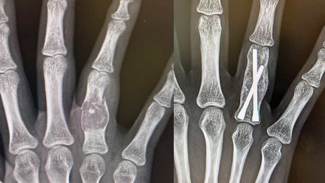
Figure 1: Limited exposure fixation of an enchondroma-associated fracture in a professional violinist, demonstrating percutaneous screw stabilization that enabled early postoperative motion.
Courtesy of Rick Tosti, MD, FAAOS
Published 6/22/2026
|
Theresa Witham
From overlooked toddler injuries to high-stakes fractures in professional athletes, hand specialists are rethinking how to match fixation choices to each patient’s goals and functional demands. Emerging implants, smarter strategies, and a focus on individualized care shaped an exploration of hand fractures during Instructional Course Lecture 163, “Fractures Hand to Fingertip,” at the AAOS 2026 Annual Meeting.
Metacarpal fixation is shifting toward intramedullary screws to support early motion
The first presentation, delivered by Omri Ayalon, MD, FAAOS, associate professor of orthopaedic surgery at New York University Grossman School of Medicine and director of the Amputation Reconstruction Center, focused on metacarpal fractures. Dr. Ayalon urged surgeons to consider both biomechanics and patient goals when considering fixation options.
Metacarpals require similar considerations to fixing femur fractures, Dr. Ayalon told AAOS Now. “In general, with a lot of fractures (and the hand is no different), we’re working to achieve length, alignment, and rotation to allow for early motion, but there’s been some exciting changes in the fixation that exists for these fractures.”
He pointed out subtle radiographic cues, among them the metacarpophalangeal (MP) joint cascade and the use of the tenodesis maneuver to assess rotation, by passively flexing and extending the wrist. “And when you extend the wrist, the fingers should typically all point towards the scaphoid tubercle,” he said. Soft-tissue stabilizers, such as the intermetacarpal ligaments, may provide greater inherent stability on the central metacarpals — an anatomical nuance that affects decisions to operate.
“When you’re trying to restore these fractures, remember that precision is more on the radial side and power is more on the ulnar side. And as such, that can impact the need to fix these fractures. When you make a fist actively, you’ll see that the fifth and the fourth [metacarpals] have more mobility, and they actually drop down in flexion. And as a result, that impacts the implications for fixing these, meaning the ulnar side of the hand can accept more deformity in these fractures and still have excellent function than on the radial side. So you’d be more likely to fix something on the radial side. Just like elsewhere in the body, it’s important to think about the morphology of the fractures and what that implies to fixation,” he said.
Although nonoperative management remains appropriate for many cases, Dr. Ayalon noted that “multiple [fractured] metacarpals in my mind, in general, is an indication for surgery, even with minimal displacement.”
His section culminated in strong advocacy for intramedullary (IM) screws, which he called “a relatively new advancement in hand surgery.” These devices, he explained, are minimally invasive, rigid enough to support immediate motion, and cosmetically appealing to patients.
He illustrated the point with a case of a 24-year-old professional basketball player. “I had to get this guy back on the court quickly. And so, he was back about three weeks later, working on his game [and] the following week, competing. He had no splint, he had a little [bandage] over the incision, and that was it. He was off to the races.”
Dr. Ayalon cautioned, however, that IM screws can be “tough to take out sometimes” and alignment must be precise. He closed his section by emphasizing, “Make sure you know what your patient’s goals are, like if they have to return to a sport or work that may impact your implant selection, as well as think about the morphology of the fracture site. And we’re always trying, in all hand fractures, to move these patients back to motion correctly.”
Two-screw constructs may reduce soft tissue trauma in proximal phalanx fractures
Next, Rick Tosti, MD, FAAOS, associate professor of orthopaedic surgery at Thomas Jefferson University and the Rothman Orthopaedic Institute, Philadelphia, presented on proximal phalanx (P1) fractures.
Dr. Tosti said he mentally divides P1 fractures into three groups: distal articular, proximal articular, and shaft fractures. Traditional K-wire constructs still work, but Dr. Tosti noted their limitations, including infection, stiff ness, and patient frustration with prolonged immobilization.
He then highlighted a minimally invasive technique that has transformed his practice: a percutaneous two-screw construct mirroring the trajectory of crossed K-wires. “The wire goes in, the cannulated drill goes over that, the screws come out, the wires come out. And now I have something that’s all internal — I can move them right away.” He added that there is “no incision,” and surgeons can choose one or two screws depending on fracture geometry.
A professional violinist’s complex enchondroma-associated fracture illustrated another application of limited-exposure fixation (Figure 1). “[We made] a very small incision over the shaft. I curetted everything and then packed it with graft, and then I put these screws in to hold it. Then I was able to get this moving within the first three days, critical for violinists,” Dr. Tosti said.
He concluded by reminding attendees, “If you can limit soft tissue trauma, I think you can also limit complications. And I think most of us can agree that early motion is superior.”
Middle phalanx and PIP fracture dislocations test even experienced surgeons
Jaehon Kim, MD, FAAOS, FACS, director of the Mount Sinai Orthopaedic Resident Research Committee and associate professor of orthopaedic surgery at the Icahn School of Medicine at Mount Sinai, New York, opened by candidly admitting, “My secret disclosure is that I’m a hand surgeon who doesn’t like fingers.” Still, his talk on middle phalanx and proximal interphalangeal (PIP) fracture dislocations tackled some of the most demanding injuries in hand trauma.
Dr. Kim reviewed common fracture patterns and emphasized the importance of rotation assessment because injuries that “may look non-displaced on X-ray… can be very, very rotated in a clinical exam.”
For oblique fractures, he cautioned against overconfidence with two-screw fixation, describing the minuscule thread diameter in small screws: “The core diameter, which is without the thread, is 1.1, and the thread diameter is 1.5, meaning the thread on each side of the screw is 0.2 millimeters. That’s a very, very small amount of thread that’s actually purchasing the bone.” Viewing the fracture from another angle — rotating the bone 90 degrees as Dr. Kim described — may reveal how little bone bridge is actually available, increasing the risk of fixation failure. Dr. Kim also demonstrated distraction-based methods — including simple wire constructs — to restore joint congruity. He noted that patients typically regain about 70 degrees of motion at the PIP joint and about 60 degrees at the distal interphalangeal (DIP) joint after treatment.
Dr. Kim emphasized that when possible, he favors “the simplest possible way” to restore stability and motion.
Carefully watch subtle pediatric injuries
Shifting to the fingertip, Kevin Chan, MD, assistant professor of orthopaedic surgery at Michigan University School of Medicine, encouraged attendees to stay alert for “snakes in the grass” when treating distal phalanx trauma. Most such fractures heal predictably nonoperatively, but exceptions can have significant consequences.
Dr. Chan reviewed tuft, shaft, and base fractures, noting that many tuft injuries heal with fibrous nonunion but function well. He emphasized examining for associated nail bed injuries — particularly in toddlers, where retained or displaced nails are common in clinic despite attempted emergency department repair.
His most impactful case was a delayed diagnosis in an 18-month-old, where a tiny fleck of bone signaled a Salter-Harris I physeal injury. “Please, please, please keep this in mind,” he urged, explaining that because the epiphysis in toddlers is still unossified cartilage, it does not appear on radiographs, making physeal injuries easy to miss in this age group. Early recognition may prevent growth disturbance and deformity.
Thumb fractures demand attention to quality of soft-tissue coverage and functional goals
Closing the session, Brandon Smetana, MD, FAAOS, associate fellowship program director and director of research at the Indiana Hand to Shoulder Center, Indianapolis, covered thumb fractures and tip injuries. Distal tip management, he emphasized, requires understanding the injury’s obliquity and whether bone is exposed.
Patients’ work demands often influence care, he said. “A lot of my patients are factory workers or farm workers and want to get back soon. So a lot of my discussion surrounds the time for that wound to contract and heal on its own, about 1 centimeter squared a month.”
Dr. Smetana detailed options for soft-tissue coverage — including VY advancement flaps, Moberg flaps, and Foucher/first dorsal metacarpal artery flaps — and offered practical tips such as using a K-wire rather than a nail bed stitch to prevent nail hooking.
For shaft fractures, he echoed the earlier speakers: “I’ve really switched to intramedullary screw technique … I think this is an optimal technique.” Thumb metacarpal fractures require careful assessment of deformity tolerance, as some patients function well despite minor malalignment. Since the thumb’s purpose is to serve as a stable post to pinch and grip against or grab larger objects, if patients “can accomplish that goal based on the clinical exam and discussion about deformities in question or expected deformities with their fracture pattern, then I will often treat those conservatively if the patient agrees,” Dr. Smetana said.
Across the session, early motion and patient-centered planning stood out
Whether discussing metacarpals, phalanges, or thumb tip injuries, the panelists consistently emphasized minimizing soft-tissue trauma, selecting implants that facilitate early motion, and tailoring choices to patient needs and goals. As Dr. Ayalon summarized, “Make sure you know what your patient’s goals are.”
Theresa Witham is managing editor for AAOS Now.