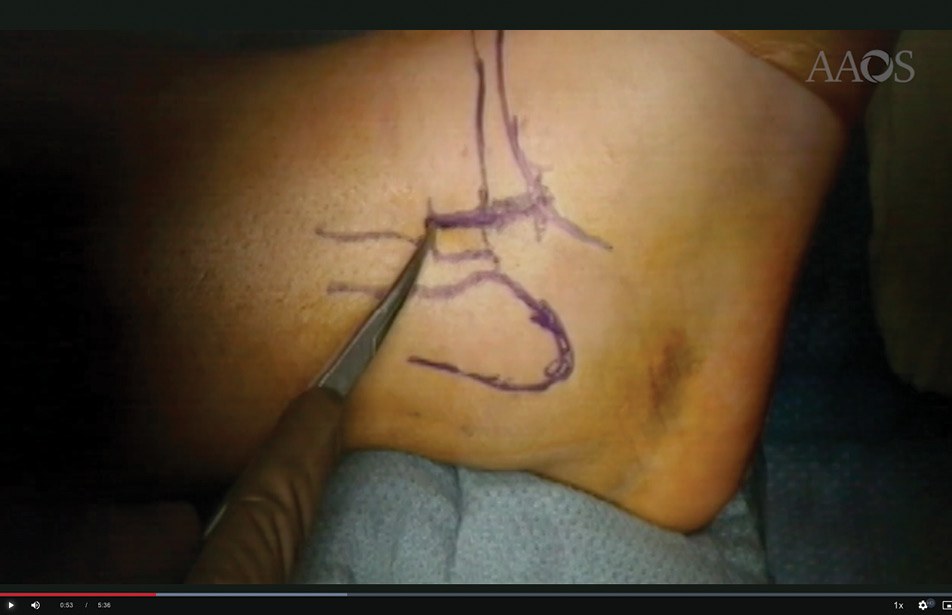
Figure 1: Skin markings outline key bony landmarks to guide a targeted approach to the syndesmosis.
Published 6/22/2026
|
Daniel R. Schlatterer, DO, MBA, FAAOS
Editor's note: The following article is a review of a video available via the AAOS Orthopaedic Video Theater (OVT). AAOS Now will routinely review “OVT Plus” videos, which are vetted by topic experts and offer CME. For more information, visit aaos.org/OVT.
Historically, syndesmotic disruptions were reduced by closed methods with percutaneous fixation if the injury pattern permitted. Closed surgical treatment largely depended on radiographic metrics to assess syndesmotic reduction based on the bony anatomy of this region. Unfortunately, closed syndesmotic malreductions have been reported as high as 50%. The reasons for malreduction are diverse and include reduction clamp placement, screw fixation trajectory, and incisura morphology.
Syndesmosis reduction directly correlates with clinical outcomes and should be accomplished by either open or closed methods. Therefore, if radiographic reduction parameters are not achieved intraoperatively via closed reduction techniques, switching to an open reduction technique is indicated to achieve reduction and maximize clinical outcomes. An open reduction may be helpful because it permits direct visualization of reduction, especially if incisura morphology is hindering the reduction.
Figure 1: Skin markings outline key bony landmarks to guide a targeted approach to the syndesmosis.
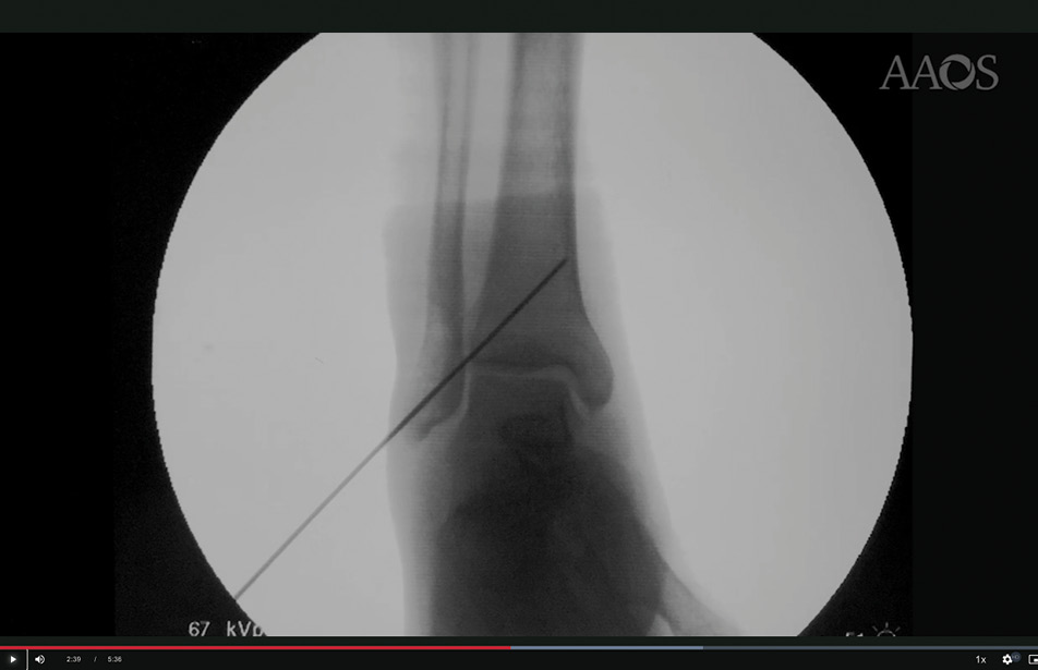
Figure 2: Intraoperative imaging demonstrates Kirschner wire placement to maintain syndesmotic reduction.
A video published in the AAOS OVT, “Limited Open Syndesmosis using Radiographs,” shows author Paul Tornetta III, MD, FAAOS, meticulously demonstrating an open reduction surgical technique. Keys parts of the technique include drawing bony landmarks on the skin (Figure 1) at the start of the syndesmotic surgery, revealing the small incision permitted by their targeted approach to the syndesmosis.
Next is the soft tissue exposure (limited joint capsule dissection). It is retracted medially and remains robust for repair at closure, which likely imparts reduction maintenance. The surgeon demonstrates placement of provisional fixation after syndesmosis reduction with a Kirschner wire (Figure 2) followed by two positional quadricortical screws, which may be key to reduction success. Also, the video states that reduction can be confirmed radiographically by restoration of the “Mercedes sign.” Finally, the provisional K-wire is removed, and the reduction is confirmed, followed by layered closure.
This surgical video offers basic instruction for surgeons interested in the open approach. Fixation options for the syndesmosis after reduction of the fibula are beyond the scope of this video. However, in terms of the number of cortices for fixation, four cortices do provide easier removal of a broken screw in the author’s experience.
While closed reduction remains an appropriate way to approach the syndesmosis, the open reduction technique permits direct inspection of the reduction and allows re-reduction if the clamp, position screw, or incisura morphology is inhibiting reduction. When a closed reduction is not achievable, consider an open technique as described by Tornetta in this OVT video.
Daniel R. Schlatterer, DO, MBA, FAAOS, is the former chair of the orthopaedic surgery residency program and former chief of orthopaedic trauma at WellStar Health System in Atlanta, Georgia. He is a member of the AAOS Now Editorial Board.
References
- Mediouni M, Schlatterer DR, Gardner M. Syndesmotic injuries: where are we now? Where do we need to go? J Foot Ankle Surg. 2017;56(5):1129-1132.
- Miller AN, Barei DP, Iaquinto JM, Ledoux WR, Beingessner DM. Iatrogenic syndesmosis malreduction via clamp and screw placement. J Orthop Trauma. 2013;27(2):100-106.
- Cherney SM, Cosgrove CT, Spraggs-Hughes AG, McAndrew CM, Ricci WM, Gardner MJ. Functional outcomes of syndesmotic injuries based on objective reduction accuracy at a minimum 1-year follow-up. J Orthop Trauma. 2018;32(1):43-51.
- Mukhopadhyay S, Metcalfe A, Guha AR, et al. Malreduction of syndesmosis—are we considering the anatomical variation? Injury. 2011;42(10):1073-1076.
- Kortekangas T, Savola O, Flinkkilä T, et al. A prospective randomised study comparing TightRope and syndesmotic screw fixation for accuracy and maintenance of syndesmotic reduction assessed with bilateral computed tomography. Injury. 2015;46(6):1119-1126.
- Miller AN, Carroll EA, Parker RJ, Boraiah S, Helfet DL, Lorich DG. Direct visualization for syndesmotic stabilization of ankle fractures. Foot Ankle Int. 2009;30(5):419-426.
- Andersen, MR, Diep LM, Frihagen, F, Castberg H, Johan MD, Madsen, JE, Figved W. Importance of syndesmotic reduction on clinical outcome after syndesmosis injuries. J Orthop Trauma. 2019;33(8):397-403. doi:10.1097/BOT.0000000000001485.
- Sagi HC, Anjan RS, Roy WS. The functional consequence of syndesmotic joint malreduction at a minimum 2-year follow-up. J Orthop Trauma. 2012;26(7):439-443.
- Cherney SM, Spraggs-Hughes AG, McAndrew CM, Ricci WM, Gardner MJ. Incisura morphology as a risk factor for syndesmotic malreduction. Foot Ankle Int. 2016;37(7):748-754.