

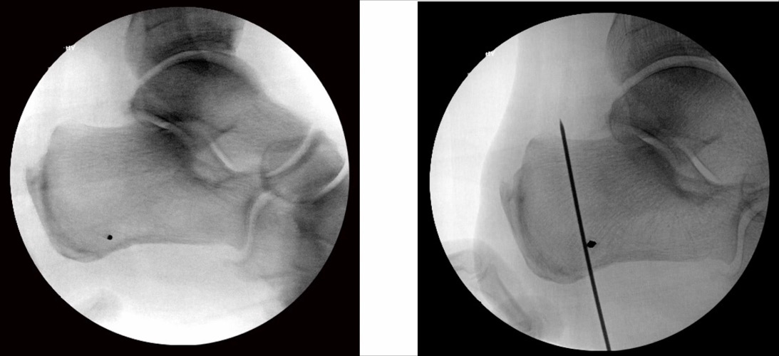
Figure 1: Intraoperative fluoroscopic images demonstrating guide pin placement at the osteotomy apex and along the planned osteotomy trajectory.
Published 9/20/2026
|
Alexandra E. Page, MD, FAAOS
At a Glance
- The Zadek osteotomy unloads the Achilles insertion.
- Percutaneous techniques may lower wound complication rates.
- Proper osteotomy planning helps avoid nerve injury.
Estimated read time: 3 minutes
Editor’s note: The following article is a review of a video available via the AAOS Orthopaedic Video Theater (OVT). AAOS Now routinely reviews OVT Plus videos, which are vetted by topic experts and offer CME. For more information, visit aaos.org/OVT.
Insertional Achilles tendinitis presents frequently, and when resistant to conservative care, surgery provides an effective solution. Risks of traditional open surgery, often performed through a posterior midline incision that releases much of the Achilles insertion, include wound complications and potential rupture at the tendon insertion. Expansion of minimally invasive surgical (MIS) techniques in the foot and ankle offers an alternative surgical option.
The Zadek osteotomy is a dorsal closing wedge of the calcaneal tuberosity, which addresses the Achilles symptoms by shortening and anteriorly rotating the tuberosity. This results in an improved mechanical environment for the Achilles, reducing tendon strain during ankle dorsiflexion and decreasing insertional impingement while also decompressing the retrocalcaneal bursa.
The Zadek osteotomy was first described in 1939 but has had a resurgence as a percutaneous method. A recent systematic review by Bakaes and colleagues reported an overall 5.8% complication rate for the percutaneous osteotomy. This contrasts with wound complications of 10% to 18% for the traditional open surgery, as shown by Hall et al.
The video “Minimally invasive Zadek osteotomy: Techniques and approach” provides a brief background on diagnosis and treatment options for insertional Achilles tendinitis but focuses on the technique overview.
Positioning is critical. The patient is placed in the lateral decubitus position with the operative foot off the table or on a radiolucent extension for imaging. A large C-arm is preferred.
Success with the percutaneous technique requires planning the osteotomy with fluoroscopy guidance. The plantar apex of the osteotomy is identified with a K-wire placed perpendicular to the lateral calcaneal wall, approximately 1 cm proximal to the plantar cortex. A second K-wire is placed percutaneously along the planned cut (Figure 1).
Through a 1-cm incision, a 3-mm high-speed bur creates the osteotomy under continuous irrigation. The authors recommend creating the vertical limb of the osteotomy, then progressively removing bone to create a 1-cm gap at the dorsal cortex. Further removal of bone from the posterior calcaneus creates the wedge, which is closed with dorsiflexion.
Fixation is obtained with percutaneously placed 7.0-mm screws. Note the text and narrative state “parallel to the osteotomy,” but the demonstrated screw placement is perpendicular. Use of headless screws can decrease the need for subsequent hardware removal.
The “safe zone” for the osteotomy is noted on a slide in the video. Talusan et al. in 2015 described the safe zone for tuberosity osteotomies as approximately 10 mm anterior to a “landmark line” defined as a line from the posterior superior apex of the calcaneal tuberosity to the origin of the plantar fascia. Surgeons should identify this zone to avoid injury to any of the multiple medial and lateral nerve branches.
With appropriate attention to technique and anatomy, the percutaneous Zadek osteotomy can offer an excellent way to address insertional Achilles tendinitis symptoms with a low complication rate.
Alexandra E. Page, MD, FAAOS, is a foot and ankle specialist in private practice in San Diego, California, and the editor-in-chief of AAOS Now.
References
- Hall S, Schipper ON, Kaplan JRM, Johnson AH, Gonzalez TA, Vulcano E. Outcomes after percutaneous Zadek osteotomy for insertional Achilles tendinopathy. Foot Ankle Int. 2024;45(9):931-939. doi:10.1177/10711007241252803.
- Hall Kiriluk S, Vulcano E, Schipper ON, et al. Percutaneous Zadek osteotomy vs open Haglund resection for insertional Achilles tendinopathy: early outcomes and complication rates. Foot Ankle Int. 2025;46(10):1103-1114. doi:10.1177/10711007251359639.
- Bakaes Y, Hall S, Jackson JB III, et al. Percutaneous vs open Zadek osteotomy for treatment of insertional Achilles tendinopathy and Haglund's deformity: a systematic review. Foot Ankle Orthop. 2024;9(2):24730114241241320. doi:10.1177/24730114241241320.
- Talusan PG, Cata E, Tan EW, Parks BG, Guyton GP. Safe zone for neural structures in medial displacement calcaneal osteotomy: a cadaveric and radiographic investigation. Foot Ankle Int. 2015;36(12):1493-1498. doi:10.1177/1071100715595696.
- Kaplan JRM, Hall S, Schipper ON, Vulcano E, Jackson JB, Gonzalez T. Percutaneous Zadek osteotomy for insertional Achilles tendinopathy and Haglund deformity: a technique tip. Foot Ankle Int. 2023;44(9):931-935. doi:10.1177/10711007231181124.
Video Details
Title: Minimally Invasive Zadek Osteotomy: Techniques and Approach
Authors: Kyle A. Lorenzo; Cary B. Chapman, MD, FAAOS; Arnaldo E. Garcia Cruz; Taylor Wingo, MD
Published: Feb. 16, 2026
Time: 7:02
Tags: Foot and Ankle, Tendon Injuries, Osteotomy, Tendons
Visit aaos.org/OVT to view this award-winning title and more than 1,600 other videos from across orthopaedic topics, institutions, practice management, and industry.