Figure 1: Guide wires placed in the supraacetabular region at the location of intended graft insertion
Published 9/10/2025
|
Ahmed Emara, MD
Editor’s note: The following article is a review of a video available via the AAOS Orthopaedic Video Theater (OVT). AAOS Now routinely reviews OVT Plus videos, which are vetted by topic experts and offer CME. For more information, visit aaos.org/OVT.
Hip dysplasia has traditionally been considered a contraindication for arthroscopic procedures, including labral repair, due to acetabular under-coverage driving labral damage. A combined approach utilizing endoscopic shelf acetabuloplasty with labral repair offers a method to address this pathology. An OVT Plus video from Soshi Uchida, MD, PhD, and colleagues detailed this technique with cam osteoplasty via periportal capsulotomy access.
Figure 1: Guide wires placed in the supraacetabular region at the location of intended graft insertion
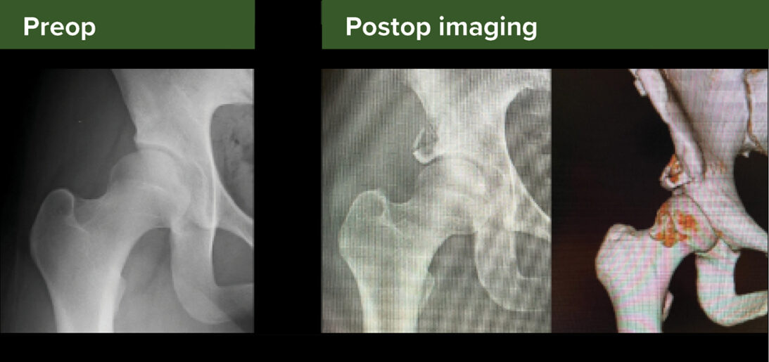
Figure 2: Preoperative versus postoperative radiographs demonstrating the outcomes of the endoscopic shelf acetabuloplasty procedure
Preparation of the shelf graft
In the video, an autologous bone graft is harvested from the ipsilateral iliac crest, ensuring the donor site is positioned 2 cm posterior to the anterior superior iliac spine to avoid lateral femoral cutaneous nerve injury.
Drill holes, 1.8 mm in diameter, are created using a Kirschner wire, with two 1.5 mm Kirschner wires inserted to guide graft placement during surgery. This critical technical step allows for adequate graft control, especially in the setting of limited arthroscopic access.
Intra-articular procedure
A 70-degree arthroscope is introduced via the anterolateral portal for visualization, followed by creation of a capsular window using a radiofrequency ablation device through the anterolateral and mid-anterior portals, which is then dilated to achieve a width of approximately 8 to 10 mm.
The arthroscope is repositioned in the anterolateral portal, and an 8 mm cannula is introduced through the mid-anterior portal. An anterosuperior labral tear, commonly identified, is repaired with suture anchors to restore labral function.
Following central compartment completion, traction is released to expose the cam lesion. Minitape is used to retract the capsule, optimizing access to the lesion. This technique allows for improved visualization and access to the full extent of the cam lesion and subsequently provides room for adequate osteoplasty. Missing this step likely increases the risk of incomplete cam resection.
Next, a motorized burr is introduced via the mid-anterior portal to perform cam osteoplasty, reshaping the femoral head-neck junction to address impingement.
Shelf acetabuloplasty procedure
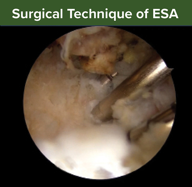
A 30-degree arthroscope is positioned in the extracapsular space for visualization. Soft tissue surrounding the reflected head of the rectus femoris is debrided with a shaver and radiofrequency ablator until clearly visible.
Four 2.3 mm guidewires are inserted with a parallel drill guide and over-drilled with a 5 mm drill to create a slot, further expanded with a shaver and osteotome (Figure 1). The slot dimensions are verified with a dilator.
The prepared graft is positioned in the slot using skewer wires and press-fit with cannulated bone tamps. Corticocancellous bone chips are packed above the shelf to ensure graft stability and promote osseous integration. Figure 2 demonstrates radiographic outcomes of this procedure.
This technique is suitable for patients with borderline or mild dysplasia, including a lateral center edge angle of 20 degrees to 25 degrees, Tönnis angle of 15 degrees or more, and a normal Shenton line. Contra-indications include severe dysplasia (lateral center edge angle <5 degrees), broken shenton line, significant femoral deformities, or osteoarthritis.></5>
Ahmed K. Emara, MD, is a senior orthopaedic surgery resident at Cleveland Clinic. Dr. Emara is chair of the AAOS Resident Assembly Education Committee and a member of the AAOS Now Editorial Board.
Video details
Title: Endoscopic shelf acetabuloplasty combined with labral repair and cam osteoplasty via periportal capsulotomy access for treating patients with acetabular dysplasia
Authors: Soshi Uchida, MD, PhD; Hokuto Fukuda, MD; Noboru Funakoshi; Yoichi Murata, MD, PhD; Hirotaka Nakashima, MD; Keisuke Nakayama, MD; Takahiro Negayama, MD; Akinori Sakai, MD, PhD; Shinichiro Takada, MD
Published: Jan. 31, 2024
Time: 12:01
Tags: Sports Medicine, Hip and Pelvis, Dysplasia
Visit aaos.org/OVT to view this title and more than 1,600 other videos from across orthopaedic topics, institutions, practice management, and industry.