

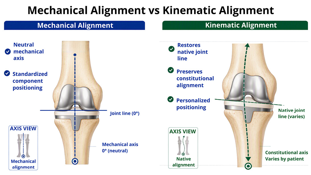
Mechanical alignment targets a neutral limb axis for all patients, whereas kinematic alignment aims to restore the patient’s pre-arthritic anatomy by preserving native joint-line orientation and constitutional alignment.
Published 10/20/2026
|
Morteza Meftah, MD; Matthew Hepinstall MD, FAAOS
At a Glance
- Kinematic alignment aims to restore a patient’s pre-arthritic anatomy.
- Evidence suggests personalized alignment may improve outcomes after UKA.
- Implant positioning is increasingly being tailored to individual knee phenotypes.
Estimated read time: 5 minutes
Unicompartmental knee arthroplasty (UKA) has long been recognized as a successful surgical solution for isolated compartment osteoarthritis. Compared to total knee arthroplasty (TKA), UKA preserves both cruciate ligaments and the unaffected compartment, yielding faster recovery and a more natural feel. Yet for decades, the native alignment of the knee has often been secondary to an alignment philosophy borrowed from TKA: restoration of a neutral mechanical axis for every patient, with bone cuts made perpendicular to the mechanical axis of the limb.
A growing body of evidence now challenges that default, suggesting that restoring a patient’s pre-arthritic anatomy, a central principle of kinematic alignment (KA), may be important for optimizing outcomes in UKA. Although tibial resection levels in conventional UKA have been influenced by medial collateral ligament (MCL) re-tensioning, the defining principle of KA is the systematic, preoperative targeting of the patient’s estimated pre-arthritic alignment, primarily through the tibial cut, restoring joint planes and accounting for cartilage loss. The application of KA philosophy to UKA is both conceptually intuitive and increasingly supported in the literature.
What is kinematic alignment?
KA is an alignment philosophy that targets restoration of a patient’s pre-arthritic joint anatomy, taking into account cartilage thickness, bony alignment, and native joint-line orientation, rather than correcting toward a standard neutral mechanical axis. In the context of UKA, this means recreating the patient’s native joint line obliquity and limb alignment, essentially resurfacing the worn compartment so that the pre-arthritic kinematics of the knee are restored. The goal is not to remove bone and cartilage to reach a predetermined angle, but rather to reconstruct the geometry that the knee had before arthritic degeneration.
KA principles are particularly relevant in UKA because the procedure inherently preserves ligamentous structures that maintain the patient’s pre-arthritic anatomy. Both cruciate ligaments remain intact, as does the collateral ligament complex. Therefore, the preserved soft tissues act as a kinematic blueprint; alignment that conflicts with that blueprint may risk abnormal loading, stiffness, and early failure.
The problem with mechanical alignment in UKA
Traditional mechanical alignment targets a postoperative hip-knee-ankle angle of 180 degrees — a straight mechanical axis from hip to ankle. While this approach has decades of precedent in TKA, its application to UKA is increasingly questioned because many patients who develop isolated medial compartment arthritis do not start with a neutral limb. Instead, they often have constitutional varus alignment.
Correcting these patients fully to neutral can shift load from the diseased medial compartment onto the lateral compartment and alter soft tissue tension across the joint. Multiple studies have shown that, in medial UKA, mild residual varus postoperatively is associated with improved outcomes compared with neutral or valgus correction. In a 2023 study published in Knee Surgery, Sports Traumatology, Arthroscopy, Bayoumi and colleagues found that restoration of pre-arthritic coronal alignment, or slight correction toward neutral relative to the pre-arthritic state, produced significantly better functional results after medial UKA.
Neutral mechanical alignment of implants may result in edge-loading when bone resections intentionally result in undercorrection of limb alignment to several degrees of varus. Furthermore, placing medial UKA tibial implants perpendicular to the mechanical axis, rather than parallel to the joint line, can result in relatively large bone resections adjacent to the tibial spines in patients with significant proximal tibia varus. These resections place greater stress on weaker cancellous bone and may predispose to fracture or implant loosening.
---
Related Content
- Alignment and Balance Methods in Total Knee Arthroplasty (JAAOS®)
- Cases Solved! Medial Unicompartmental Knee Arthroplasty (AAOS resource)-
---
The CPAK framework: quantifying pre-arthritic anatomy
The introduction of the Coronal Plane Alignment of the Knee (CPAK) classification has helped identify knee coronal phenotypes in a systematic way. CPAK uses standing radiographs to categorize knees into nine phenotypes based on two parameters: the arithmetic hip-knee-ankle angle (aHKA), which estimates constitutional limb alignment, and joint line obliquity (JLO), which reflects how oblique the native joint line is relative to the ground.
In the context of UKA without bone loss, CPAK offers a systematic framework to estimate a patient’s pre-arthritic knee alignment before loss of cartilage. The CPAK classification therefore provides a reproducible, radiograph-based target for personalized alignment. In UKA, preservation of native coronal alignment has also been associated with superior patient-reported outcomes, supporting the idea that alignment should be matched to the patient’s constitutional phenotype rather than forced toward a universal neutral axis.
A mild postoperative varus for medial UKA and mild postoperative valgus for lateral UKA tend empirically to produce favorable outcomes, consistent with the constitutional alignment patterns commonly seen in patients with isolated compartment disease.
KA vs MA: literature review
A 2024 retrospective study from MedStar Washington Hospital Center directly compared kinematically and mechanically aligned UKAs using the Oxford Knee Score (OKS) and Forgotten Joint Score (FJS). The kinematic alignment group demonstrated better scores on both outcome measures, with a statistically significant improvement in OKS. CPAK-based radiographic analysis also confirmed systematic differences between the alignment philosophies, particularly in lateral distal femoral angle. A systematic review of medial UKA found that kinematic alignment appears safe, with no increase in revision or complication risk when performed within appropriate boundaries.
Although a systematic review of total knee arthroplasty reported improved patient satisfaction and joint awareness with kinematic alignment without increased revision risk, direct UKA-specific randomized data remain limited. Prospective randomized controlled trials comparing KA and MA specifically in UKA would be required to establish higher-level evidence, but may not be appropriate given the available evidence that the best outcomes in medial UKA come from re-tensioning the MCL and leaving the limb in slight varus — essentially by restoring pre-arthritic alignment.
Practical implications
Transitioning to a KA philosophy in UKA does not require a radical departure from current practice. It begins with preoperative planning: reviewing standing full-length radiographs to characterize the patient’s CPAK phenotype and estimate pre-arthritic alignment. Several software platforms and robotic-assisted systems now integrate these calculations into surgical workflows, allowing surgeons to plan component position relative to the patient’s own anatomy rather than a population-averaged neutral.
As robotic-assisted UKA becomes more widespread, personalized alignment is likely to become easier to execute, with greater precision and reproducibility. Haptic robotic platforms allow surgeons to preoperatively plan component position relative to the patient’s own phenotype, then execute bone cuts within a defined kinematic target zone in real time. A 2025 technique description introduced functional positioning for robotic medial UKA, a complementary personalization concept that targets congruent femorotibial contact and balanced soft tissue tension throughout the arc of motion rather than a fixed coronal target alone, reflecting the broader shift toward individualized, kinematically informed component placement in UKA.
Conclusion
Kinematic alignment represents a conceptual refinement in UKA that returns the focus to what the procedure was always intended to accomplish: restoring the patient’s pre-arthritic anatomy. A growing body of evidence supports the view that targeting constitutional alignment of the limb and the implants, rather than a universal mechanical neutral, is associated with better functional outcomes and higher patient satisfaction without an increase in revision risk. The ideal alignment thresholds continue to be refined. Nevertheless, as our tools for quantifying pre-arthritic anatomy improve and robotic platforms make personalized alignment more reproducible, kinematic alignment in UKA is poised to evolve from an emerging philosophy into an increasingly validated alignment strategy.
Morteza Meftah, MD, is an associate professor of orthopaedic surgery and a member of the Division of Adult Reconstructive Surgery at NYU Langone Orthopedic Hospital, where his clinical and research focus is total and unicompartmental knee arthroplasty, robotic-assisted surgery, personalized alignment, and patient-reported outcomes.
Matthew Hepinstall MD, FAAOS, is a board-certified orthopaedic hip and knee surgeon with expertise in robot-assisted and less invasive surgical techniques including partial knee replacement and anterior hip replacement. He is an associate professor of orthopaedic surgery at NYU Langone Health in New York.
References
- Bayoumi T, Burger JA, Ruderman LV, et al. Restoration or relative overcorrection of pre-arthritic coronal alignment leads to improved results following medial unicompartmental knee arthroplasty. Knee Surg Sports Traumatol Arthrosc. 2023;31(9):3981-3991. doi:10.1007/s00167-023-07441-9.
- McEwen P, Omar A, Hiranaka T. Unicompartmental knee arthroplasty: what is the optimal alignment correction to achieve success? The role of kinematic alignment. J ISAKOS. 2024;9(6):100334. doi:10.1016/j.jisako.2024.100334.
- Rivière C, Sivaloganathan S, Villet L, et al. Kinematic alignment of medial UKA is safe: a systematic review. Knee Surg Sports Traumatol Arthrosc. 2022;30(3):1082-1094. doi:10.1007/s00167-021-06462-6.
- MacDessi SJ, Griffiths-Jones W, Harris IA, Bellemans J, Chen DB. Coronal Plane Alignment of the Knee (CPAK) classification. Bone Joint J. 2021;103-B(2):329-337. doi:10.1302/0301-620X.103B2.BJJ-2020-1050.R1.
- Kim SE, Yun KR, Lee JM, Lee MC, Han HS. Preserving coronal knee alignment of the knee (CPAK) in unicompartmental knee arthroplasty correlates with superior patient-reported outcomes. Knee Surg Relat Res. 2024;36:1. doi:10.1186/s43019-023-00204-3.
- Digennaro V, Ferri R, Panciera A, et al. Coronal plane alignment of the knee (CPAK) classification and its impact on medial unicompartmental knee arthroplasty: exposing an unexpected external shift of limb mechanical axis in case of prearthritic constitutional valgus alignment: a retrospective radiographic study. Knee Surg Relat Res. 2024;36:14. doi:10.1186/s43019-024-00217-6.
- Gulati J, Fernandez C, Ricci J, et al. A retrospective comparison of mechanically vs. kinematically aligned unicompartmental knee arthroplasties. J Orthop. 2024;52:33-36. doi:10.1016/j.jor.2024.02.016.
- Andriollo L, Mazzella GG, Koutserimpas C, et al. Functional positioning in robotic medial unicompartmental knee arthroplasty: a step-by-step technique. SICOT-J. 2025;11:34. doi:10.1051/sicotj/2025028.