

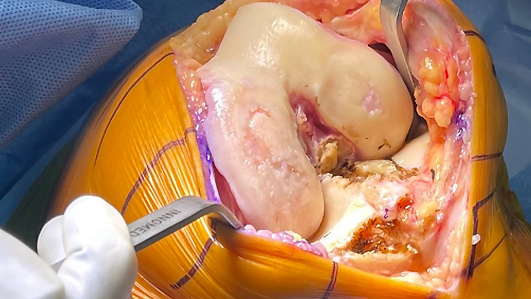
Intraoperative inspection revealed a small full-thickness cartilage defect in the lateral weight-bearing compartment despite the absence of obvious lateral compartment disease on preoperative radiographs. Because the lateral compartment was no longer preserved, the planned unicompartmental knee arthroplasty (UKA) was converted to total knee arthroplasty (TKA).
Courtesy of Peter Gold, MD
Published 10/19/2026
|
Peter Gold, MD
At a Glance
- Shared decision making helps identify appropriate candidates for UKA and TKA.
- Many traditional contraindications to UKA have evolved with modern implants and robotics.
- Final procedure selection may depend on intraoperative assessment of cartilage and compartment involvement.
Estimated read time: 5 minutes
As modern surgical treatment of knee osteoarthritis (OA) has evolved, my practice has developed a two-tiered system for indicating knee arthroplasty. The first decision involves identifying properly indicated candidates in which moderate to severe osteoarthritis significantly affects quality of life in the setting of failed conservative treatment (medications, exercise, injections, etc.). The second tier focuses on the pathology pattern and identifying which knee compartments are affected by disease.
This second tier frames the discussion of unicompartmental (UKA) versus total knee arthroplasty (TKA) with the patient and leads to a final decision based on intraoperative findings. This article provides a practical approach to identifying UKA versus TKA candidates, developing shared decision making with the patient, and making the final intraoperative decision.
Expand the indications for UKA
Indications for UKA have expanded since the Kozinn and Scott criteria from 1989. Early restrictions on location of pain, anterior cruciate ligament (ACL) stability, weight, deformity/contractures, and age were due to poor fixation and balancing techniques of older implants, reliance on manual instrumentation, and limits in polyethylene quality. Berend and Lombardi tested newer indications in 2007, allowing for use in younger patients and obese patients and the discovery that patients with patellofemoral disease and preoperative anterior knee pain still had successful outcomes. With the use of robotics in UKA, indications have continued to expand with success in alignment accuracy, better balancing techniques, prevention of overcorrection, and rotational precision.
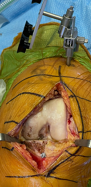
Direct visualization confirms isolated medial compartment disease with an intact lateral compartment and lateral trochlea. These findings support proceeding with medial unicompartmental knee arthroplasty (UKA) rather than total knee arthroplasty (TKA).
Courtesy of Peter Gold, MD
Intraoperative inspection revealed a small full-thickness cartilage defect in the lateral weight-bearing compartment despite the absence of obvious lateral compartment disease on preoperative radiographs. Because the lateral compartment was no longer preserved, the planned unicompartmental knee arthroplasty (UKA) was converted to total knee arthroplasty (TKA).
Courtesy of Peter Gold, MD
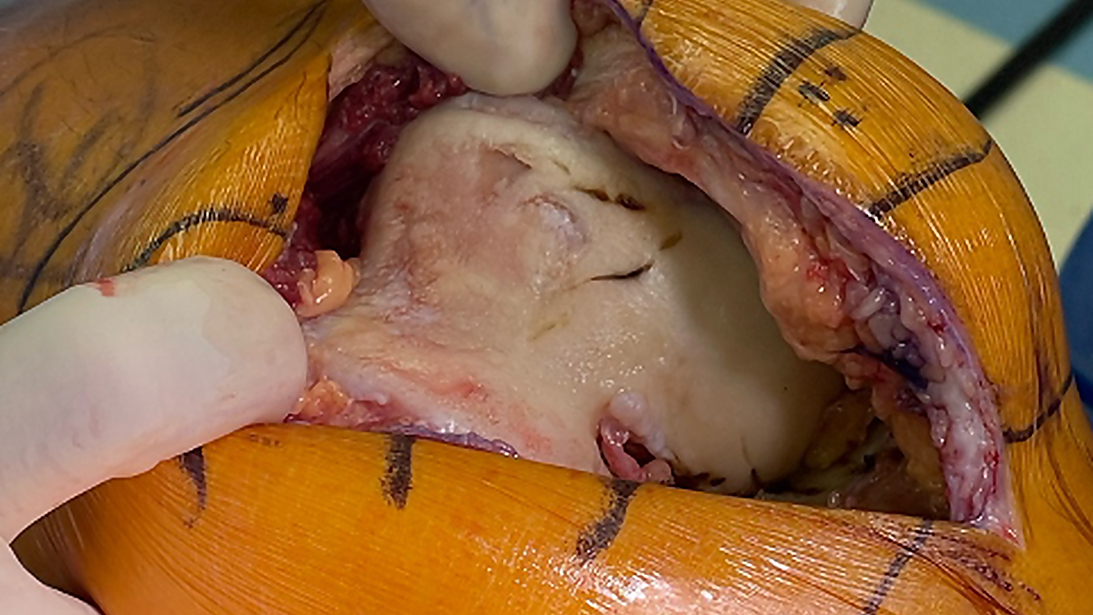
Patellofemoral disease limited to the medial or central trochlea may be considered for patellofemoral arthroplasty. In this case, the disease extends into the lateral trochlea, making the patient unsuitable for medial unicompartmental arthroplasty and necessitating conversion to total knee arthroplasty (TKA).
Courtesy of Peter Gold, MD
Evaluate the arthroplasty candidate
In my practice, clinical assessment is identical for both UKA and TKA candidates. First, it is essential to listen to the patient’s story, understanding duration, location, and how much their day-to-day or quality of life is affected by their knee arthritis. Next, I focus on identifying all nonoperative treatments the patient has tried and how effective those treatments have been. This should be coupled with reviewing radiographs with the patient, typically a weight-bearing series: AP, Rosenberg/flexion PA, lateral, and sunrise/merchant view.
Throughout this review, the patient should be educated on distinguishing three weight-bearing compartments of the knee (medial, lateral, and patellofemoral). Radiographs should reveal which of the three appears to be affected with signs of moderate to severe OA (joint space narrowing, osteophyte formation, and bone sclerosis). If a patient’s radiographic knee OA is limited to one compartment, they should be considered a candidate for UKA.
As I transition to the physical exam, I find it important to note the patient’s functional arc of motion, flexion contractures, and passively correctable deformities. Severe deformities or flexion contractures may make UKA a less favorable option. Prior teaching held that pain location should match radiographic findings; however, pain location correlates poorly with radiographic and intraoperative findings in osteoarthritis, and preoperative pain location does not predict outcome after UKA, likely because unicompartmental disease produces effusion and synovitis that refer pain nonspecifically. Localized pain should therefore not be a prerequisite.
Shared decision making: UKA or TKA?
Shared decision making with patients then moves to the two tiers. In tier-one discussions, the patient has moderate to severe osteoarthritis that is affecting quality of life despite exhausting nonoperative treatment measures. The second-tier discussion begins with patients whose radiographs display moderate to severe osteoarthritic changes in only one weight-bearing compartment and have favorable physical exam findings. These patients can be presented with the option for a UKA versus a TKA, with the caveat that the actual decision on which procedure will be performed is made intraoperatively, with direct visualization of the articular cartilage in all three compartments.
Benefits and risks of UKA are honestly shared. The benefits include faster functional recovery, maintaining more native knee kinematics, preserving unaffected anatomy, a higher Forgotten Joint Score, greater satisfaction performing functional activities, higher satisfaction rates, and ACL and bone preservation. Risks include higher revision rates in registry data, although conversion of a failed UKA to TKA carries fewer complications than aseptic revision TKA (while still underperforming a primary), and the possibility of OA progression in the remaining compartments. TKA is described as durable and addresses all compartments, with substantially better long-term registry survivorship, although the same tricompartmental resection that provides that durability sacrifices the cruciates and alters native kinematics, and revision TKA is more morbid than conversion of a UKA. Setting proper expectations about recovery and longevity is important.
---
Related Content
- Personalized alignment in UKA: Moving beyond mechanical neutral (AAOS Now)
- Can robotics expand the use of unicompartmental knee arthroplasty? (AAOS Now)
- Medial unicompartmental arthroplasty of the knee (JAAOS®)
---
Making the final intraoperative decision
My appropriate patients are then consented to both UKA versus TKA in every single case. It must be clear that decisions are not made preoperatively but only on intraoperative joint visualization and confirmation of pattern of knee OA pathology. The patients must be comfortable with you making the final decision to do the least amount of surgery needed but everything the knee requires.
In practice, the final determination is made after surgical exposure. Following the arthrotomy and minimal medial release, all three compartments are inspected directly. The patellofemoral compartment is evaluated, and the lateral compartment and lateral meniscus are assessed through a full range of motion before committing to UKA. For efficiency, the operative team is initially set up for a partial knee procedure, with all TKA instrumentation and trays available in the operating room. If intraoperative findings indicate that TKA is more appropriate, the team begins opening the total knee trays while the surgeon completes the exposure and preparation of the knee. When robotics or navigation are used, preplanning for both UKA and TKA can further streamline the transition and avoid delays.
One caveat is a bias toward ordering a preoperative MRI for patients with potential lateral unicompartmental disease to try to limit the amount of intraoperative conversion from a lateral UKA to TKA via the lateral parapatellar approach. Even with favorable MRI findings preoperatively, the surgeon must be comfortable doing a TKA through a lateral approach if intraoperative findings contradict MRI imaging.
This shared decision process, and specifically the intraoperative decision, is revisited with the patient in the preoperative holding area, where we explain that during the approach we will expose all three compartments of the knee for direct visualization of the articular cartilage and contralateral meniscus. To move forward with a medial or lateral UKA, the contralateral side must show no signs of cartilage damage (partial or full thickness) through a full range of motion (must take the knee into deep flexion to see posterior condyle), as well as an intact meniscus on the contralateral side. Signs of borderline cartilage thinning should be probed to confirm cartilage stability. Any sloughing or concern about unhealthy cartilage during probing would prompt conversion to a TKA.
With emerging data, I find greater leeway in assessing the patellofemoral compartment than previously taught, with the relevant threshold being bone loss and grooving on the lateral side of the joint. Medial UKA remains indicated in patients with medial trochlear, central trochlear, or patellar cartilage defects, however severe, provided the lateral side is preserved. The need for a stable ACL may no longer be required for a UKA if you are using a fixed bearing device and the patient does not report significant instability preoperatively. The operative team should be ready for your final intraoperative decision; once made, the proper trays for either UKA or TKA are opened. This can be done simultaneously while the surgeon finishes the approach and knee exposure without adding operative time.
With expanding indications and a stepwise approach, perhaps more knee arthroplasty patients can be UKA candidates. Patients with radiographic unicompartmental disease may be considered partial versus total knee candidates, and preoperative shared decision making should be discussed. Ultimately, intraoperative findings determine the appropriate candidates for UKA versus TKA.
Peter Gold, MD, is an orthopaedic surgeon at the Panorama Orthopedic & Spine Center in Golden, Colorado.
References
- Kozinn SC, Scott R. Unicondylar knee arthroplasty. J Bone Joint Surg Am. 1989;71(1):145–150.
- Berend KR, Lombardi AV Jr, Adams JB. Obesity, young age, patellofemoral disease, and anterior knee pain: identifying the unicondylar arthroplasty patient in the United States. Orthopedics 2007;30(5 Suppl):19.
- Bell SW, Anthony I, Jones B, MacLean A, Rowe P, Blyth M. Improved accuracy of component positioning with robotic-assisted unicompartmental knee arthroplasty: data from a prospective, randomized controlled study. J Bone Joint Surg Am 2016;98(8):627. https://doi.org/10.2106/JBJS.15.00664.
- Liddle AD, Pandit H, Jenkins C, et al. Preoperative pain location is a poor predictor of outcome after Oxford unicompartmental knee arthroplasty at 1 and 5 years. Knee Surg Sports Traumatol Arthrosc 2013;21(11):2421-2426. https://doi.org/10.1007/s00167-012-2211-3.
- Hamilton TW, Pandit HG, Maurer DG, et al. Anterior knee pain and evidence of osteoarthritis of the patellofemoral joint should not be considered contraindications to mobile-bearing unicompartmental knee arthroplasty: a 15-year follow-up. Bone Joint J 2017;99-B(5):632-639. https://doi.org/10.1302/0301-620X.99B5.BJJ-2016-0695.R2.
- Liddle AD, Pandit H, Judge A, Murray DW. Patient-reported outcomes after total and unicompartmental knee arthroplasty: a study of 14,076 matched patients from the National Joint Registry for England and Wales. Bone Joint J 2015;97-B(6):793-801. https://doi.org/10.1302/0301-620X.97B6.35155.
- Brilliant ZR, Garvey MD, Haffner R, Chiu YF, Mayman DJ, Blevins JL. Unicompartmental knee arthroplasty patients have lower joint awareness and higher function at 5 years compared to total knee arthroplasties: a matched comparison. J Arthroplasty 2023;38(8):1464-1470. https://doi.org/10.1016/j.arth.2023.01.063.
- Patil S, Colwell CW Jr, Ezzet KA, D’Lima DD. Can normal knee kinematics be restored with unicompartmental knee replacement? J Bone Joint Surg Am 2005;87(2):332-338. https://doi.org/10.2106/JBJS.C.01467.
- Liddle AD, Pandit H, Judge A, Murray DW. Optimal usage of unicompartmental knee arthroplasty: a study of 41,986 cases from the National Joint Registry for England and Wales. Bone Joint J 2015;97-B(11):1506-1511. https://doi.org/10.1302/0301-620X.97B11.35551.
- Forlenza EM, Burnett RA, Acuña AJ, Levine BR, Gililland JM, Della Valle CJ. Revision unicompartmental knee arthroplasty: worse than a primary, but better than a revision total knee arthroplasty. J Arthroplasty. 2025;40(1 Suppl):S297-S303. https://doi.org/10.1016/j.arth.2024.12.026.
- Liddle AD, Judge A, Pandit H, Murray DW. Adverse outcomes after total and unicompartmental knee replacement in 101,330 matched patients: a study of data from the National Joint Registry for England and Wales. Lancet 2014;384(9952):1437-1445. https://doi.org/10.1016/S0140-6736(14)60419-0.
- Evans JT, Walker RW, Evans JP, Blom AW, Sayers A, Whitehouse MR. How long does a knee replacement last? A systematic review and meta-analysis of case series and national registry reports with more than 15 years of follow-up. Lancet 2019;393(10172):655-663. https://doi.org/10.1016/S0140-6736(18)32531-5.
- Plancher KD, Briggs KK, Tucker EE, et al. The role of severe lateral facet patellar osteoarthritis in patient selection for success of a medial unicompartmental knee arthroplasty: mean follow-up of 10 years. J Arthroplasty. 2023;38(7 Suppl 2):S145-S149.
- https://doi.org/10.1016/j.arth.2023.05.024.