

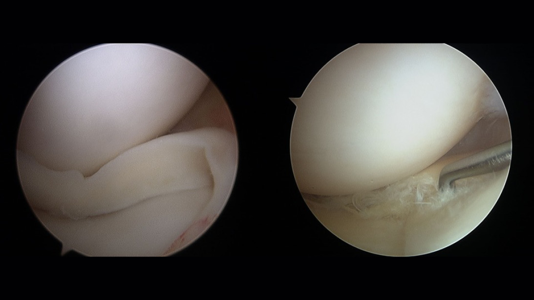
Figure 1: (a) Bucket handle tear of the medial meniscus. (b) Complex tear of the white-white zone of the medial meniscus.
Source: Stone JA, Salzler MJ, Parker DA, Becker R, Harner CD. Degenerative meniscus tears - assimilation of evidence and consensus statements across three continents: state of the art. J ISAKOS. 2017;2(2):108-119. doi:10.1136/jisakos-2015-000003. Figures 1 and 2. Used under CC BY-NC-ND license.
Published 11/19/2026
|
Alireza Nazemi, MD, MS; David Swanson, MD; Ben Joseph, MD
At a Glance
- Meniscal preservation through repair should be the primary goal when tear morphology and patient factors permit.
- Tear location, size, pattern, and tissue quality are critical determinants in the repair versus meniscectomy decision.
- Long-term outcomes favor repair over meniscectomy in appropriate candidates, with implications for early degenerative changes and functional preservation.
Estimated read time: 6 minutes
Editor’s note: This article is part of “Orthopaedic Fundamentals,” a recurring AAOS Now series highlighting core principles widely embraced across the orthopaedic community. The series examines ideas that can improve efficiency and outcomes within and outside the OR, informing training surgeons as well as providing practical refreshers for experienced surgeons.
Meniscal tears represent one of the most common knee injuries encountered, present in up to one third of all knee MRIs in adult patients. About half of these tears may become symptomatic in nature. When symptoms fail to resolve with conservative management, the tear pattern blocks motion, or patients have significant pain or mechanical symptoms, surgery may be indicated.
In these cases, the decision between meniscus repair and partial meniscectomy has significant implications for patient outcomes and long-term joint health. Meniscus preservation has emerged as a fundamental principle in knee preservation, as meniscectomy accelerates degenerative changes regardless of tear pattern or patient age. Not all tears are repairable, and the surgeon should develop a systematic approach to identifying which tears can be successfully repaired and which lend themselves to meniscectomy for pain relief and functional restoration.
Figure 1: (a) Bucket handle tear of the medial meniscus. (b) Complex tear of the white-white zone of the medial meniscus.
Source: Stone JA, Salzler MJ, Parker DA, Becker R, Harner CD. Degenerative meniscus tears - assimilation of evidence and consensus statements across three continents: state of the art. J ISAKOS. 2017;2(2):108-119. doi:10.1136/jisakos-2015-000003. Figures 1 and 2. Used under CC BY-NC-ND license.
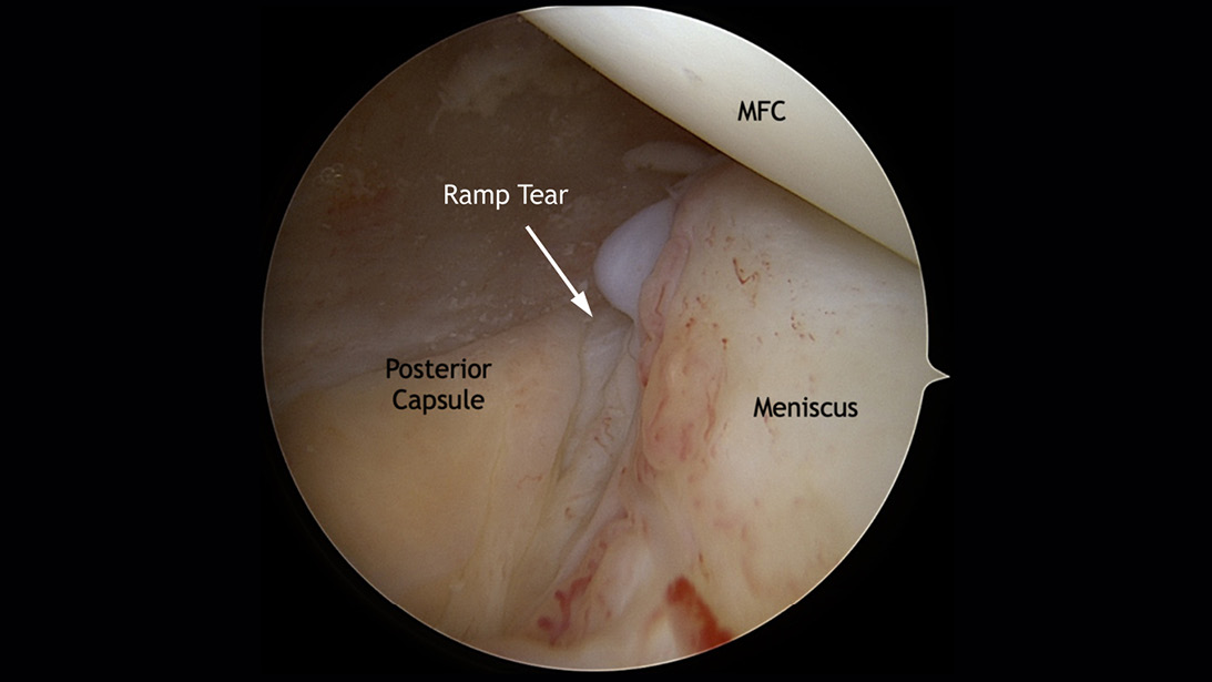
Figure 2: Ramp lesion viewed by a Gillquist maneuver.
Source: Tollefson LV, Tuca MJ, Tapasvi S, LaPrade RF. Medial meniscus ramp tears: state of the art. J ISAKOS. 2025;10:100380. doi:10.1016/j.jisako.2024.100380. Figure 5. Used under CC BY-NC-ND license.
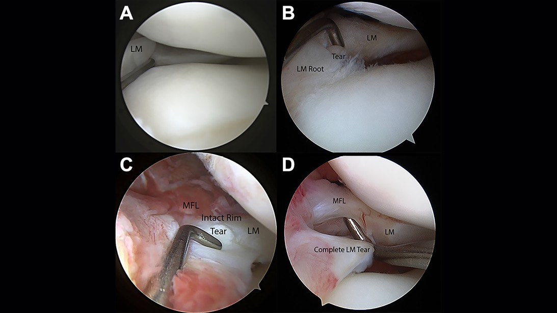
Figure 3: Arthroscopic views of the four types of posterior horn lateral meniscal oblique radial tears. A Type 1: partial oblique tear <10 mm from the lateral root. B Type 2: complete oblique tear <10 mm from the root attachment, not involving the root. C Type 3: incomplete oblique tear extending >10 mm from the root. D Type 4: complete oblique tear extending >10 mm from the root.
Source: Krych AJ, LaPrade MD, Cook CS, Leland D, Keyt LK, Stuart MJ, Smith PA. Lateral meniscal oblique radial tears are common with ACL injury: a classification system based on arthroscopic tear patterns in 600 consecutive patients. Orthop J Sports Med. 2020;8(5):2325967120921737. doi:10.1177/2325967120921737. Figure 2. Used under CC BY-NC license.
Pearl: Preoperative assessment guides repair candidacy; intraoperative findings and tissue quality determine final management.
Why preserve the meniscus?
The meniscus plays critical roles in load distribution, shock absorption, proprioception, and joint stability. Meniscectomy may disrupt these functions and accelerate degenerative changes, with the amount of degeneration proportionate to the size of the removed fragment. Long-term outcomes following meniscectomy reveal significantly higher rates of osteoarthritis, particularly in younger patients, regardless of tear morphology or preexisting cartilage health.
Epidemiological evidence demonstrates that meniscectomy confers twice the risk of radiographic osteoarthritis compared to meniscal repair in matched cohorts. Beyond structural joint changes, patients undergoing meniscectomy report higher rates of functional decline, persistent knee pain, and activity limitations compared to repair patients. These findings favor meniscal preservation when repair is technically feasible and tissue quality supports healing.
---
Related Content
- Orthopaedic Fundamentals: Rotator cuff repair evaluation, visualization, and diagnostic arthroscopy (AAOS Now)
- Study identifies predictors and patterns of concomitant meniscus injury in ACL reconstruction patients (AAOS Now)
- Meniscus Repair Part 1: Biology, Function, Tear Morphology, and Special Considerations (JAAOS®)
---
Tear morphology and repairability assessment
Tear pattern, location, and size are primary determinants of repairability. The meniscus is conventionally divided into the vascular (red-red and red-white zones) and avascular (white-white zone) regions. Tears within 6 mm of the meniscal capsule (red-red and red-white zones) have excellent healing potential with blood supply and fibrocartilage formation and are more amenable to repair (Figure 1a). In contrast, tears isolated to the central avascular region lack intrinsic blood supply and have lower healing rates — relying on synovial fluid for nourishment — and should generally not be repaired (Figure 1b).
Tear morphology also influences repair success. Longitudinal or bucket-handle tears in the peripheral meniscus represent ideal repair candidates, with healing rates exceeding 90% when vascular tissue is present and repair is performed within six months of the tear. Radial tears extending from the inner margin are increasingly recognized as repairable, particularly in younger patients and peripheral tears. Horizontal cleavage tears may be amenable to repair, but in patients with existing degenerative changes this may not be the case.
In the setting of ACL injury, ramp lesions and lateral meniscus oblique radial tears (LMORTs) should be identified. Ramp lesions should be identified by a Gillquist maneuver and possible use of a posteromedial portal for repair (Figure 2). LMORTs may require meniscectomy, root repair, or side-to-side repair based on tear pattern. A careful examination of the lateral meniscus posterior horn and root will determine the best treatment (Figure 3).
Critical patient factors in repair decision-making
Beyond tear morphology, several patient and tissue factors guide the repair versus meniscectomy decision. Patient age, tear chronicity, associated injuries, and baseline activity level all contribute to repair candidacy. Younger patients with acute injuries represent optimal repair candidates and should undergo repair whenever feasible to maximize longevity of meniscal tissue and delay osteoarthritis onset.
Pearl: Tear chronicity, location, and tissue quality are intraoperative determinants; preoperative imaging provides guidance but does not replace direct visualization.
Meniscal repair techniques and healing optimization
Modern meniscal repair techniques include inside-out (suture passage through the posterior capsule), all-inside (suture anchors deployed from the articular surface), and outside-in approaches. Root repairs are also commonly performed when indicated.
Inside-out repair is widely utilized for peripheral longitudinal tears, offering excellent anatomic reduction and healing rates exceeding 90% in appropriately selected cases. All-inside repair techniques have expanded indications, particularly for more central tears, LMORTs, and posterior horn medial meniscus tears. Outside-in techniques are particularly helpful in repairing anterior horn tears. Studies show that inside-out repairs have lower failure rates for medial meniscus bucket handle tears, and all-inside repairs may have lower neurovascular injury rates at the expense of potential implant-related complications. An additional downside to all-inside techniques is the cost of devices themselves, as several are often needed for a complete repair. It is the authors’ view that the decision regarding repair technique should factor in tear pattern, location, and surgeon experience.
Healing optimization involves several key principles: early repair within weeks of injury (versus chronic tears), restoration of meniscal vascularity and hemostasis at the tear site, preservation of meniscal substance, and adherence to postoperative protected weight-bearing protocols. Biologic enhancement strategies — including platelet-rich plasma (PRP), bone marrow aspirate concentrate (BMAC), and fibrin clot scaffolding — have been shown to improve healing rates.
When partial meniscectomy remains appropriate
Partial meniscectomy remains indicated when repair is not feasible or fails. Specific scenarios include avascular tears in the central white-white zone with no vascular supply, complex tears with multiple fragmentation patterns, and degenerative horizontal cleavage tears. Weight-bearing radiographs, particularly Rosenberg views, help assess joint space collapse when considering meniscectomy for degenerative tears. Meniscectomy is also appropriate for symptomatic meniscal cysts, displaced bucket-handle tears with poor tissue quality or white-white zone fragments, and in the setting of failed prior meniscal repairs.
When meniscectomy is selected, a conservative resection approach that preserves as much viable meniscal tissue as possible is preferred over aggressive resection. Extensive meniscectomy should be avoided, as total or near-total meniscectomy substantially increases osteoarthritis risk and long-term morbidity. Surgeon technique during meniscectomy — including preservation of the peripheral meniscus, careful debridement of only torn fragments, and properly visualizing the tear — influences functional outcomes.
Pearl: Repair should be attempted in peripheral tears with adequate tissue quality; meniscectomy is reserved for avascular, symptomatic degenerative, or irreparable tears.
Long-term outcomes and shared decision making
Long-term follow-up studies demonstrate superior outcomes with meniscal repair compared to meniscectomy. At 10-year follow-up, repair patients have higher functional scores, greater activity participation, and lower radiographic osteoarthritis progression. Despite the need for potentially longer operative times and more complex rehabilitation protocols, repair represents a durable, cost-effective strategy when technically feasible. With the advent of newer repair techniques, certain types of meniscal repairs have become more efficient and timely. This is particularly appealing in more complicated tears or knees with other associated injuries and will hopefully be one less deterrent to choosing repair in the future.
Shared decision making with the patient is essential. Patients should understand that repair may require up to a six-week period of protected weight bearing (PWB) and 12 weeks until complete meniscal healing and carry a 15% re-tear rate depending on tear pattern — with root and radial tear repairs having a longer PWB period than longitudinal tear repairs. In contrast, meniscectomy provides immediate symptomatic relief and quicker return to unrestricted activity but sacrifices meniscal tissue and predisposes to long-term degenerative changes. In younger, active patients with repairable tears, the case for repair is compelling. In older patients with complex tears or advanced degenerative changes, meniscectomy may be more appropriate.
The decision between meniscal repair and partial meniscectomy represents a critical juncture in knee preservation. By systematically assessing tear morphology, tissue quality, patient factors, and expected outcomes, surgeons can optimize meniscal preservation in appropriate candidates while acknowledging that some tears require meniscectomy. This nuanced, individualized approach — informed by current evidence and explicit patient communication — represents the standard practice in orthopaedic surgery.
Alireza Nazemi, MD, MS, is a fellowship-trained orthopaedic surgeon with a specialized focus on sports medicine and arthroscopic surgery, practicing in California. David Swanson, MD, is a fellowship-trained orthopaedic surgeon with a specialized focus on sports medicine and arthroscopic surgery, practicing in New York.
Ben Joseph, MD, is a fellowship-trained orthopaedic surgeon with a specialized focus on sports medicine and arthroscopic surgery, practicing in Florida.