

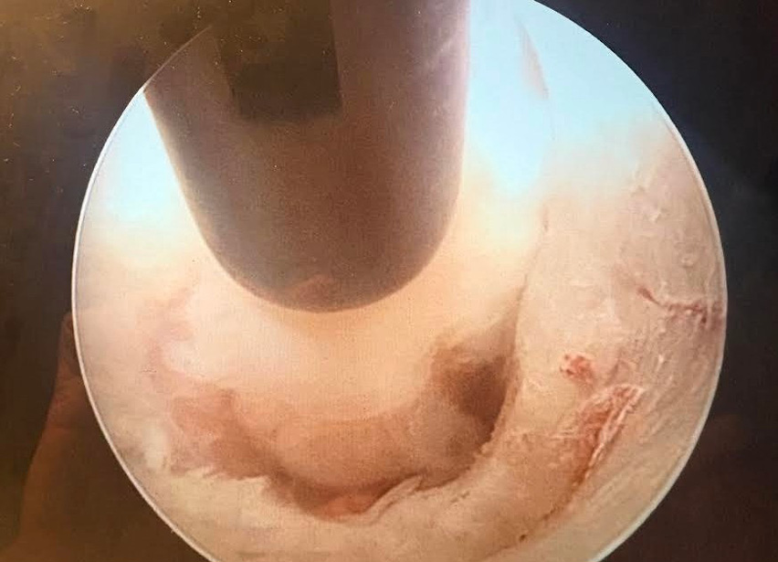
Endoscopic view of the neural foramen during posterior full-endoscopic cervical foraminotomy and diskectomy. The image demonstrates the working corridor established after bony decompression, allowing direct visualization of the decompressed neural elements.
Source: OVT Posterior Full-Endoscopic Cervical Foraminotomy and Diskectomy: Surgical Techniques and Review of Outcomes
Published 10/20/2026
|
Robert Kamil, MD, MBA; Paul S. Issack, MD, PhD, FAAOS, FACS
At a Glance
- PECF can address unilateral cervical radiculopathy from foraminal stenosis.
- The technique uses a systematic six-step approach to decompression.
- Careful identification of anatomic landmarks is critical to safe execution.
Estimated read time: 3 minutes
Editor’s note: The following article is a review of a video available via the AAOS Orthopaedic Video Theater (OVT). AAOS Now routinely reviews OVT Plus videos, which are vetted by topic experts and offer CME. For more information, visit aaos.org/OVT .
Posterior full-endoscopic cervical foraminotomy and diskectomy (PECF) is indicated for patients with unilateral cervical radiculopathy secondary to foraminal stenosis caused by soft-disc herniation or uncovertebral hypertrophy after failure of nonoperative management. In the OVT presentation “Posterior Full-Endoscopic Cervical Foraminotomy and Diskectomy: Surgical Techniques and Review of Outcomes,” Sang Hun Lee, MD, PhD, demonstrates a six-step technique for neuroforaminal decompression occurring from both soft-disc and hard-disc pathology. Contraindications include central or paracentral disc herniation medial to the uncovertebral joint, central canal stenosis, myelopathy, segmental instability, and predominant axial neck pain.
The procedure begins with fluoroscopic localization of the interpedicular space followed by docking of the working cannula at the facet-laminar junction. Bipolar radiofrequency dissection removes the paravertebral soft tissues until the inferior articular process (IAP), superior articular process (SAP), facet joint overlap point, and lateral ligamentum flavum are clearly identified. Identification of these landmarks establishes the working corridor and defines the boundaries of the subsequent bony decompression. Facet resection proceeds in a sequential fashion as in open surgery. The inferomedial corner of the IAP is resected with an endoscopic burr until the medial SAP and ligamentum flavum insertion are exposed. The SAP is then drilled to enlarge the neural foramen while maintaining anatomic orientation. The exiting nerve root remains protected beneath the ventral facet capsule, epidural loose soft tissue, and longitudinal epidural veins until deliberate exposure is performed. These tissues are subsequently removed with bipolar radiofrequency and endoscopic forceps to expose the compressed nerve root (Figure 1).
---
Related Content
- Experts discuss the promises, challenges of biologics in spine care (AAOS Now)
- Intervertebral Disk Degeneration and Emerging Biologic Treatments (JAAOS®)
---
For extruded soft-disc herniations, an endoscopic dissector is advanced caudal to the nerve root and rotated in the cranial direction to expose the disc fragment, which typically compresses the nerve from a caudal-to-cranial direction. Herniated fragments are removed with endoscopic graspers, followed by systematic exploration of both the cranial and caudal recesses to identify retained fragments. A nerve hook is then used to confirm a freely mobile, tension-free nerve root without residual compression. It may also be necessary to approach disc fragments cranial to the nerve root.
The video also demonstrates decompression of combined soft-disc and uncovertebral osteophyte pathology. Following adequate foraminotomy, the nerve root is protected while an endoscopic burr followed by osteotomes removes the compressing uncovertebral osteophyte. When additional working space is required, limited resection of the superomedial aspect of the inferior pedicle facilitates access to the osteophyte while maintaining continuous visualization of the nerve root.
The video provides a systematic demonstration of PECF and will be of great interest to spine fellows and spine surgeons exploring endoscopic spinal approaches.
Robert Kamil, MD, MBA, is an orthopaedic house officer at New York–Presbyterian/Lower Manhattan Hospital.
Paul S. Issack, MD, PhD, FAAOS, FACS, is a clinical associate professor in the Department of Orthopaedic Surgery, Weill Cornell Medical College, and a trauma and adult reconstruction orthopaedic surgeon at New York–Presbyterian/Lower Manhattan Hospital. He is also a member of the AAOS Now Editorial Board.
Video Details
Title: Posterior Full-Endoscopic Cervical Foraminotomy and Diskectomy: Surgical Techniques and Review of Outcomes
Author: Sang Hun Lee, MD, PhD
Published: Jan. 23, 2026
Time: 6:18
Tags: Spine, Cervical, Endoscope